Two Cases of Uveal Effusion Syndrome
- Affiliations
-
- 1The Institute of Vision Research, Department of Ophthalmology, Yonsei University College of Medicine, Seoul, Korea. semekim@yumc.yonsei.ac.kr
- KMID: 1099048
- DOI: http://doi.org/10.3341/kjo.2006.20.2.124
Abstract
- PURPOSE: To report a case of uveal effusion syndrome associated with hypotony and a case of uveal effusion syndrome in nanophthalmos. METHODS: The first case was a 25-year-old man who presented with decreased visual acuity in the left eye and hypotony. Fundus examination revealed choroidal effusion and retinal detachment with a thickened eyeball. Partial thickness sclerotomy and sclerectomy were performed. The second case was a 13-year-old boy who had uveal effusion syndrome with a nanophthalmic eye. RESULTS: In the patient with hypotony, intraocular pressure was well maintained following partial thickness sclerotomy and sclerectomy, and choroidal effusion and retinal detachment were reduced. The visual acuity of the nanophthalmic patient was well maintained during a 3-year follow-up period without treatment. CONCLUSIONS: appropriate treatment modalities should be considered depending on the ophthalmic condition of the individual patient.
Keyword
MeSH Terms
Figure
-
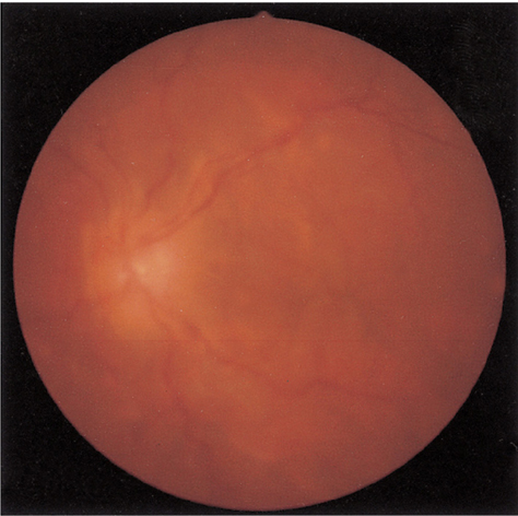
Fig. 1 Preoperative color photograph of case 1 showing shallow retinal detachments and choroidal folding in the left eye.

Fig. 2 B-scan ultrasound in case 1 showed thickened eyeball and choroidal detachment in the left eye.

Fig. 3 Color photograph of case 1 at postoperative 2 months showing attached retina and near complete resolution of choroidal detachment.

Fig. 4 Color photograph of case 2 showing serous retinal detachments and epiretinal membrane.
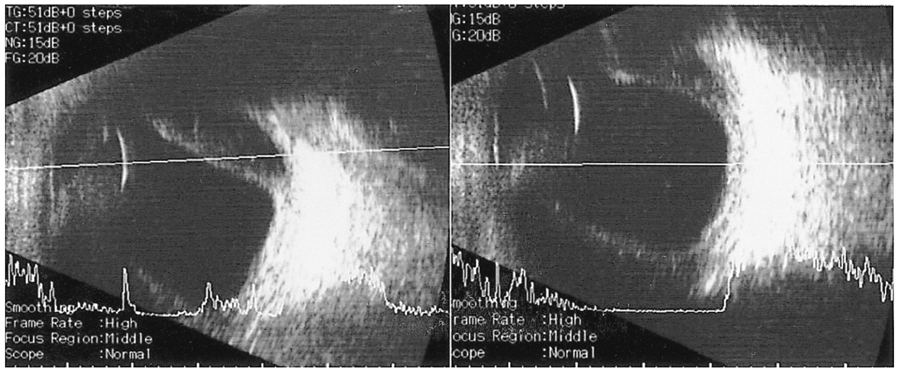
Fig. 5 B-scan ultrasound in case 2 showing thickened eyeball, peripheral retinal detachment and choroidal detachment.
Cited by 1 articles
-
Protective Effect of Preoperative Intraocular Pressure Reduction on Corneal Endothelium in Cataract Surgery
Yong Sun Ahn, Yang Kyung Cho
J Korean Ophthalmol Soc. 2015;56(4):521-531. doi: 10.3341/jkos.2015.56.4.521.
Reference
-
1. Schepens CL, Brockhurst RJ. Uveal effusion. I. Clinical picture. Arch Ophthalmol. 1963. 70:189–201.2. Brockhurst RJ. Nanophthalmos with uveal effusion: a new clinical entity. Arch Ophthalmol. 1975. 93:1989–1999.3. Brockhurst RJ. Vortex vein decompression for nanophthalmic uveal effusion. Arch Ophthalmol. 1980. 98:1987–1990.4. Gass JDM. Uveal effusion syndrome: a new hypothesis concerning pathogenesis and technique of surgical treatment. Trans Am Ophthalmol Soc. 1983. 81:246–260.5. Trelstad RL, Silbermann NN, Brockhurst RJ. Nanophthalmic sclera: ultrastructural, histochemical, and biochemical observations. Arch Ophthalmol. 1982. 100:1935–1938.6. Stewart DH III, Streeten BW, Brockhurst RJ, et al. Abnormal scleral collagen in nanophthalmos: an ultrastructural study. Arch Ophthalmol. 1991. 109:1017–1025.7. Uyama M, Takahashi K, Kozaki J, et al. Uveal effusion syndrome: clinical features, surgical treatment, histologic examination of the sclera, and pathophysiology. Ophthalmology. 2000. 107:441–449.8. Young RD. The ultrastructural organization of proteoglycans and collagen in human and rabbit scleral matrix. J Cell Sci. 1985. 74:95–104.9. Oum BS. Uveal effusion. J Korean Ophthalmol Soc. 1986. 27:1013–1017.10. Song SW, Kwak NH, Huh W. A case of nanophthalmos. J Korean Ophthalmol Soc. 1996. 37:687–692.
