Optical Coherence Tomographic Finding in a Case of Macular Coloboma
- Affiliations
-
- 1Department of Ophthalmology, Seoul National University College of Medicine, Seoul, Korea. jiani4@snu.ac.kr
- 2Seoul National University Bundang Hospital, Seongnam, Korea.
- 3Seoul Artificial Eye Center, Seoul National University Hospital Clinical Research Institute, Seoul, Korea.
- KMID: 1099036
- DOI: http://doi.org/10.3341/kjo.2007.21.3.175
Abstract
- PURPOSE: To report the optical coherence tomography (OCT) findings in a patient with unilateral macular coloboma. METHODS: A 12-year-old male was presented with macular coloboma in the left eye. The optical coherence tomography was performed with fluorescein angiography (FA). RESULTS: The OCT revealed the crater-like depression in the macula, demonstrating atrophic neurosensory retina, and an absence of retinal pigment epithelium and choroid in the lesion. FA showed hypofluorescence corresponding to the size of the lesion in both early and late frames without leakage of dye at any stage. CONCLUSIONS: The OCT can be beneficial to confirm the diagnosis of macular coloboma.
MeSH Terms
Figure
-
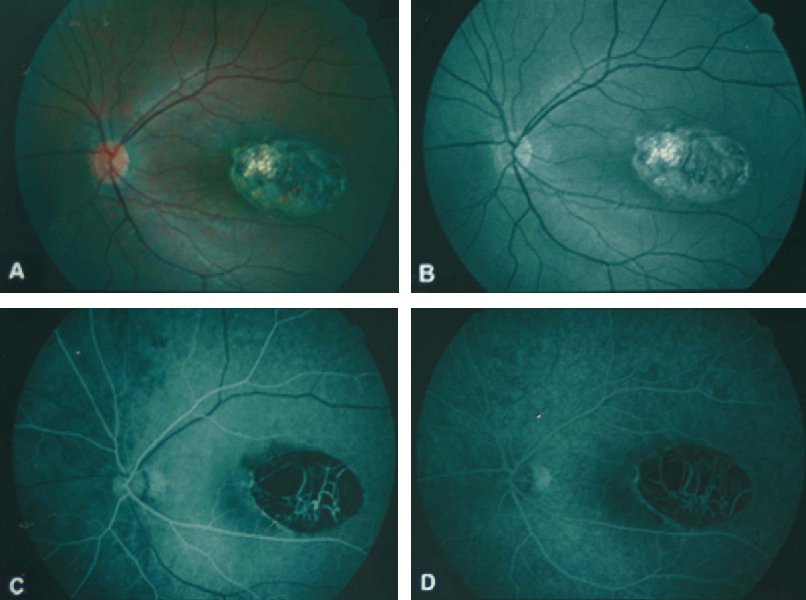
Fig. 1 Color fundus photography shows a sharply-demarcated, oval-shaped, excavated lesion involving the temporal half of the macula (A). Red-free retinal nerve fiber layer photography shows intact papillomacular bundle around the lesion (B). Large choroidal vessels and bared sclera are observed at the base. Fluorescence angiography shows hypofluorescence corresponding to the size of the lesion in both early (C) and late frames (D) without leakage of dye.
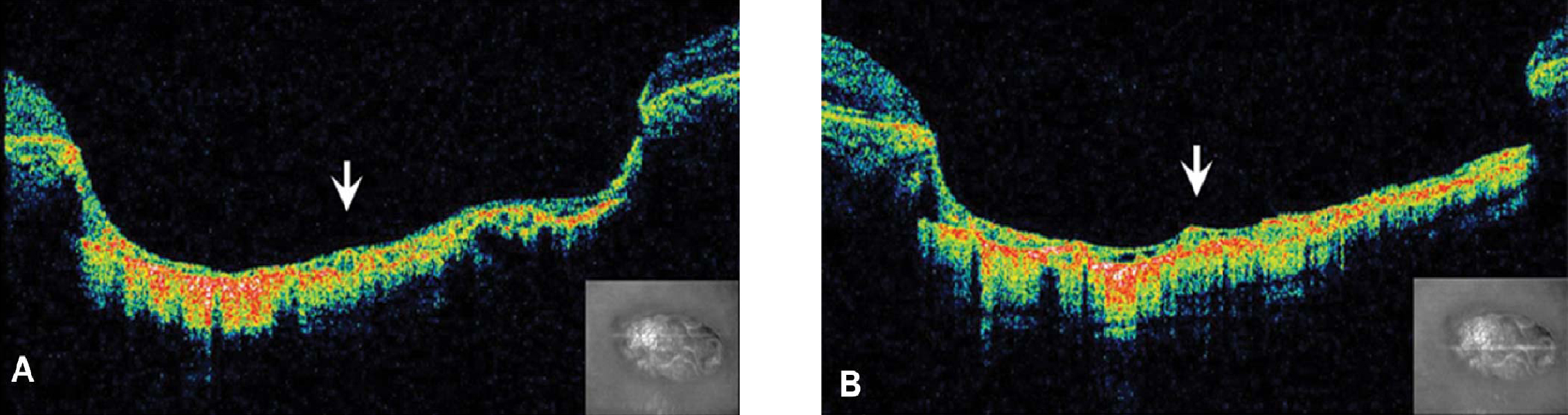
Fig. 2 Optical coherence tomography shows that the neurosensory retina is abnormally atrophied, and retinal pigment epithelium and choroid are absent in the lesion (A). A thin membranous structure of atrophied neurosensory retina is outlining the bare sclera and large choroidal vessels (B).
Reference
-
1. Duke-Elder S, Cook C. Duke-Elder S, Cook C, editors. Congenital deformities. System of Ophthalmology. 1963. v. 6. St. Louis: Mosby Company;chap. 14.2. Chen MS, Yang CH, Huang JS. Bilateral macular coloboma and pigmented paravenous retinochoroidal atrophy. Br J Ophthalmol. 1992. 76:250–251.3. Satorre J, Lopez JM, Martinez J, Pinera P. Dominant macular colobomata. J Pediatr Ophthalmol Strabismus. 1990. 27:148–152.4. Pian D, Ferrucci S, Anderson SF, Wu C. Paramacular Coloboma. Optom Vis Sci. 2003. 80:556–563.5. Sharma S, Naqvi A, Cruess AF. Bilateral macular colobomatas. Can J Ophthalmol. 1996. 31:27–28.
- Full Text Links
-
- Actions
-
Cited
- CITED
-
- Close
- Share
-
- Similar articles
-
- Two cases of congenital macular coloboma
- Patterns of Macular Edema in Patients with Branch Retinal Vein Occlusion on Optical Coherence Tomography
- The Correlation between Visual Acuity and Patterns of Diabetic Macular Edema in OCT Images
- A Case of Macular Edema after Rosiglitazone Use
- Optical Coherence Tomographic Evaluation of Impending Idiopathic Macular Hole Repaired by Vitrectomy
