Korean J Ophthalmol.
2007 Sep;21(3):137-141. 10.3341/kjo.2007.21.3.137.
A Comparison of the Efficacy of Cataract Surgery Using Aqualase(R) with Phacoemulsification Using MicroFlow(R) System
- Affiliations
-
- 1Department of Ophthalmology and Visual Science, Kangnam St. Mary's Hospital, College of Medicine, The Catholic University of Korea, Seoul, Korea.ckjoo@catholic.ac.kr
- 2Korean Eye Tissue and Gene Bank Related to Blindness, Seoul, Korea.
- KMID: 1099027
- DOI: http://doi.org/10.3341/kjo.2007.21.3.137
Abstract
- PURPOSE: To compare the outcomes after phacoemulsification performed with the AquaLase(R) and phacoemulsification in MicroFlow(R) system, including surgically induced astigmatism (SIA), corneal endothelial cell damage and postoperative recovery of visual acuity. METHODS: The cataracts of Lens Opacities Classification System, version III (LOCS III) nuclear grade below 2 were subjected in this study. Nineteen eyes underwent cataract operation using AquaLase(R) (Alcon Laboratories, Fort Worth, Texas, U.S.A.). A control group (19 eyes) used the MicroFlow(R) system (Millenium, Stortz, U.S.A.) and was selected by matching age, sex, systemic disease, corneal astigmatism and corneal endothelial cell density. All the surgeries were performed by the same operator. SIA, corneal endothelial cell loss, visual acuity, and corneal thickness were evaluated postoperatively. RESULTS: SIA in the group using AquaLase(R) was less than that of the group using MicroFlow(R) system (P=0.022) at 2 months postoperatively. Evaluation of corneal endothelial cell loss, recovery of visual acuity and corneal thickness found no statistically significant differences between the two groups. CONCLUSIONS: Cataract surgery using AquaLase(R) induces less surgically induced astigmatism in mild to moderate cataracts.
MeSH Terms
Figure
-
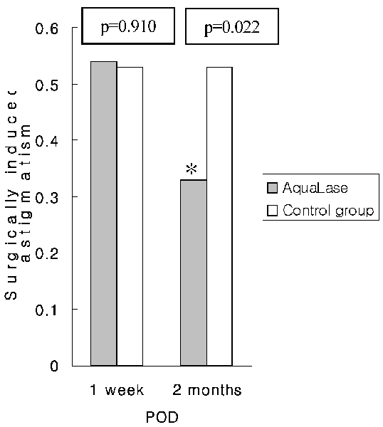
Fig. 1 Mean surgically induced astigmatism after operation. (*: statistically significant difference)

Fig. 2 Endothelial cell loss (A), endothelial cell density (B), coefficient of variation of cell area (C), and percentage of hexagonal cells (D) in the AquaLase® group and control group at 2 months postoperatively. No statistically significant differences were found between the two groups.
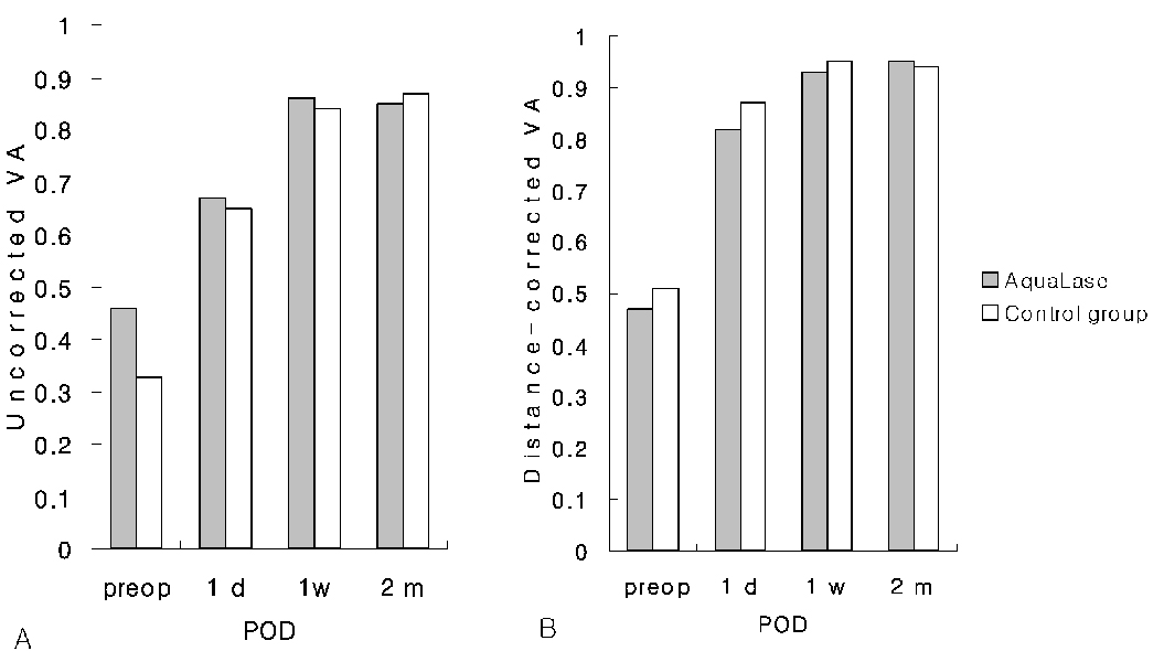
Fig. 3 Uncorrected visual acuity (VA) (A) and distance-corrected visual acuity (b) between AquaLase® group and control group after operation. There were no statistically significant differences between the two groups (p>0.05).
Reference
-
1. Mackool RJ, Brint SF. AquaLase: a new technology for cataract extraction. Curr Opin Ophthalmol. 2004. 15:40–43.2. Lehmann RP. Surgeon:AquaLase lens system improves cataract removal. Ocular surgery news. 2003. 21:July 15.3. Chylack LT Jr, Wolfe JK, Singer DM, et al. The Lens Opacities Classification System, Version III (LOCS III). Arch Ophthalmol. 1993. 107:991–997.4. Yanoff M, Duker JS. Ophthalmology. 2004. 2nd ed. St. Louis: Mosby;229–230.5. Sandoval HP, Sarraf OA, Vroman DT, Solomon KD. Corneal endothelial cell damage after lens extraction using the fluid-based system compared to ultrasound phacoemulsification in human cadaver eyes. Cornea. 2004. 23:720–722.6. Lundberg B, Jonsson M, Behndig A. Postoperative corneal swelling correlates strongly to corneal endotheial cell loss after phacoemulsification cataract surgery. Am J Ophthalmol. 2005. 139:1035–1041.7. Hayashi K, Hayashi H, Nakao F, Hayashi F. Risk factors for corneal endothelial injury during phacoemulsification. J Cataract Refract Surg. 1996. 22:1079–1084.8. Walkow T, Anders N, Klebe S. Endothelial cell loss after phacoemulsification: relation to preoperative and intraoperative parameters. J Cataract Refract Surg. 2000. 26:727–732.9. Bourne RR, Minassian DC, Dart JK, et al. Effect of cataract surgery on the corneal endothelium : modern phacoemulsification compared with extracapsular cataract surgery. Ophthalmology. 2004. 111:679–685.10. Bilinska E, Wesolek-Czernik A, Synder A, Omukecki W. Surgically induced astigmatism after cataract phacoemulsification. Klin Oczna. 2004. 106:756–759.11. George R, Rupauliha P, Sripriya AV, et al. Comparison of endothelial cell loss and surgically induced astigmatism following conventional extracapsular cataract surgery, manual small-incision surgery and phacoemulsification. Ophthalmic Epidemiol. 2005. 12:293–297.12. Alio J, Rodriguez-Prats JL, Galal A, Ramzy M. Outcomes of microincision cataract surgery versus coaxial phacoemulsification. Ophthalmology. 2005. 112:1997–2003.13. Huang FC, Tseng SH. Comparison of surgically induced astigmatism after sutureless temporal clear corneal and scleral frown incision. J Cataract Refract Surg. 1998. 24:477–481.14. Sugar A, Schertzer RM. Clinical course of phacoemulsification wound burns. J Cataract Refract Surg. 1999. 25:688–692.15. Donnenfeld ED, Olson RJ, Solomon R, et al. Efficacy and wound-termperature gradient of WhiteStar phacoemulsification through a 1.2 mm incision. J Cataract Refract Surg. 2003. 29:1097–1100.16. Sporl E, Genth U, Schmalfuss K, Seiler T. Thermomechanical behavior of the cornea. Ger J Ophthalmol. 1996. 5:322–327.17. Sippel KC, Pineda R Jr. Phacoemulsification and thermal wound injury. Semin Ophthalmol. 2002. 17:102–109.
- Full Text Links
-
- Actions
-
Cited
- CITED
-
- Close
- Share
-
- Similar articles
-
- The Effect of Phacoemulsification with a Liquefaction Device on Cornea Endothelium
- The Relationship Between the Density of Lens and Liquefaction Time Using Liquefaction Device
- Comparison of Effective Phacoemulsification Time between Femtosecond Laser-Assisted Cataract Surgery and Conventional Cataract Surgery
- The Comparison of Postoperative Astigmatic Changes Between Phscoemulsification and Planned Extracapsular Cataract Extraction
- The Quadri: Combined Phototherapeutic Keratectomy, Penetrating Keratoplasty, Phacoemulsification and Posterior Chamber Lens Implantation
