The Effects of Optic Disc Factors on Retinal Nerve Fiber Layer Thickness Measurement in Children
- Affiliations
-
- 1Department of Ophthalmology, College of Medicine, Dongsan Medical Center, Keimyung University, Daegu, Korea. lsy3379@dsmc.or.kr
- KMID: 1099006
- DOI: http://doi.org/10.3341/kjo.2008.22.2.115
Abstract
- PURPOSE: We analyzed the effect of the changes of the optic disc area (ODA) caused by the axial length and the refractive error, and the consequent changes of the distance from the optic disc margin to the circular scan (OD-CS) of Optical coherence tomography (OCT) on the measurement of the retinal nerve fiber layer thickness(RNFLT) were examined. METHODS: One hundred two eyes of 51 children (age range 4 to 15 years) were measured using OCT including the RNFLT. For the ODA and the OD-CS, the relative area formed by the ODA and the circular scan was obtained. In addition, the correlation of the refractive error and the axial length to the optic disc factors was assessed. RESULTS: As hyperopia progresses to myopia, the axial length became longer, the ODA became smaller (r=-0.442, p=0.000) and the OD-CS showed a tendency to increase (r=0.471, p=0.000). As the OD-CS became longer, the measured average RNFLT decreased significantly (r=-0.248, p=0.012), and the ODA and the OD-CS showed a significant correlation to the RNFL thickness that was measured in the nasal and inferior areas, the S2, N2 and N3 areas and the I1 area. CONCLUSIONS: As ODA becomes smaller and the OD-CS becomes longer, the RNFLT measured in the nasal and inferior areas, the S2, N2, N3, I1 area has a tendency to be thinner. Hence, consideration of the disc area is required when interpreting the RNFLT of these eyes.
MeSH Terms
Figure
-
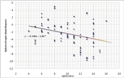
Fig. 1 Relationship between the spherical equivalent (diopter) and age (years). Note the relationship showing a decrease of the spherical equivalent with increasing age.
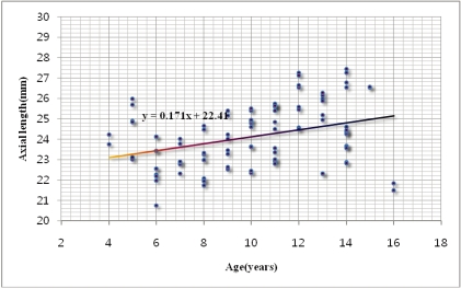
Fig. 2 Relationship between axial length (mm) and age (years). Note the relationship showing an increase of the axia length with increasing age.
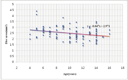
Fig. 3 Relationship between the optic disc diameter (mm2) and age (years). Note the relationship showing a decrease of the optic disc diameter with increasing age.
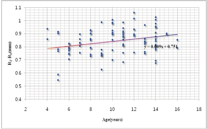
Fig. 4 Relationship between the distance from the optic disc margin to the circular scan of optical coherence tomography (R2-R1) and age (years). Note the relationship showing an increased of the distance with increasing age.
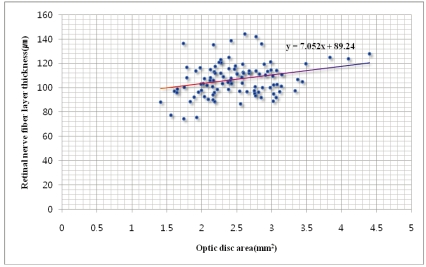
Fig. 5 Relationship between the optic disc area (mm2) and the retinal nerve fiber layer thickness (µm). Note the significant statistical correlation of the increased retinal nerve fiber layer thickness with the increasing disc diameter, and between the optic disc diameter and the retinal nerve fiber layer thickness.
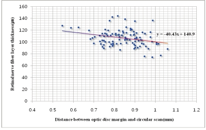
Fig. 6 Relationship between the distance from the optic disc margin to the circular scan of optical coherence tomography (mm) and the retinal nerve fiber layer thickness (µm). Note the significant statistical correlation of the increased retinal nerve fiber layer thickness with the increasing distance, and between the distance and the retinal nerve fiber layer thickness.
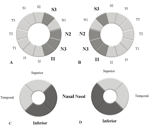
Fig. 7 (A, B) 12 equal 30-degree sectors of the retinal nerve fiber layer thickness that was measured by a circular scanning diameter of 3.4 mm around the optic disc by optical coherence tomography in the right and left eye, The dark colored sectors have significant statistical correlation between the retinal nerve fiber layer thickness and the optic disc area. (C,D) Four 90 degree quadrants of the retinal nerve fiber layer thickness that were measured by a circular scan. The dark colored quadrants have significant correlation between the retinal nerve fiber layer thickness and the optic disc area.
Reference
-
1. Schuman JS, Pedut-Kloizman T, Hertmark E, et al. Reproducibility of nerve fiber layer thickness measurements using optical coherence tomography. Ophthalmology. 1996; 103:1889–1898. PMID: 8942887.
Article2. Jones AL, Sheen NJ, North RV, Morgan JE. The Humphrey optical coherence tomography scanner: quantative analysis and reproducibility study of the normal human retinal nerve fiber layer. Br J Ophthalmol. 2001; 85:673–677. PMID: 11371486.3. Blumenthal EZ, Williams JM, Weinreb RN, et al. Reproducibility of nerve fiber layer thickness measurements by use of optical coherence tomography. Ophthalmology. 2000; 107:2278–2282. PMID: 11097610.4. Carpineto P, Ciancaglini M, Zuppardi E, et al. Reliability of nerve fiber layer thickness measurements using optical coherence tomography in normal and glaucomatous eyes. Ophthalmology. 2003; 110:190–195. PMID: 12511365.
Article5. Salchow DJ, Oleynikov YS, Chiang MF, et al. Retinal nerve fiber layer thickness in normal children measured with optical coherence tomography. Ophthalmology. 2006; 113:786–791. PMID: 16650674.
Article6. Huynh SC, Wang XY, Rochtchina E, Mitchell P. Peripapillary retinal nerve fiber layer thickness in a population of 6-year-old children: findings by optical coherence tomography. Ophthalmology. 2006; 113:1583–1592. PMID: 16949443.7. Paunescu LA, Schuman JS, Price LL, et al. Reproducibility of nerve fiber thickness, macular thickness, and optic nerve head measurements using StratusOCT. Invest Ophthalmol Vis Sci. 2004; 45:1716–1724. PMID: 15161831.
Article8. Quigley HA, Coleman AL, Dorman-Pease ME. Larger optic nerve heads have more nerve fibers in normal monkey eyes. Arch Ophthalmol. 1991; 109:1441–1443. PMID: 1929937.
Article9. Jonas J.B., Schmidt AM, Muller-Bergh JA, et al. Human optic nerve fiber count and optic disc size. Invest Ophthalmol Vis Sci. 1992; 33:2012–2018. PMID: 1582806.10. Caprioli J, Miller JM. Optic disc rim area is related to disc size in normal subjects. Arch Ophthalmol. 1987; 105:1683–1685. PMID: 3689192.
Article11. Budenz DL, Anderson DR, Varma R, et al. Determinants of normal retinal nerve fiber layer thickness measured by Stratus OCT. Ophthalmology. 2007; 114:1046–1052. PMID: 17210181.
Article12. Alamouti B, Funk J. Retinal thickness decreases with age: an OCT study. Br J Ophthalmol. 2003; 87:899–901. PMID: 12812895.
Article13. Choi SW, Lee SJ. Thickness changes in the fovea and peripapillary retinal nerve fiber layer depend on the degree of myopia. Korean J Ophthalmol. 2006; 20:215–219. PMID: 17302206.
Article14. Hoh ST, Greenfield DS, Mistlberger A, et al. Optical coherence tomography and scanning laser polarimetry in normal, ocular hypertensive, and glaucomatous eyes. Am J Ophthalmol. 2000; 129:129–135. PMID: 10682963.
Article15. HA SW, Rho SH. Age-related differences of optical coherence tomography data in Koreans. J Korean Ophthalmol Soc. 2005; 46:2037–2044.16. Song JH, Kim E, Yoo JM. Analysis of RNFL thickness and optic nerve head measured with OCT in children. J Korean Ophthalmol Soc. 2007; 48(10):1346–1353.
Article17. Johnson BM, Miao M, Sadun AA. Age-related decline of human optic nerve axon populations. Age. 1987; 20:5–9.
Article18. Asrani S, Zou S, d'Anna S, et al. Noninvasive mapping of the normal retinal thickness at the posterior pole. Ophthalmology. 1999; 106:269–273. PMID: 9951475.19. Jung Hee-Jin, Hyun Jae Hoon, Kim Young Il, et al. Normal macular thickness measured macular mapping of OCT3. J Korean Ophthalmol Soc. 2004; 45:962–968.20. Mikelberg FS, Yidegiligne HM, White VA, Schulzer M. Relation between optic nerve axon diameter to scleral canal area. Ophthalmology. 1991; 98:60–63. PMID: 2023734.21. Varma R, Skaf M, Barron E. Retinal nerve fiber layer thickness in normal human eyes. Ophthalmology. 1996; 103:2114–2119. PMID: 9003346.
Article22. Park SE, Choi KR. Analysis of optic disc size and retinal nerve fiber thickness. J Korean Ophthalmol Soc. 2002; 43:395–401.23. Leung CK, Mohamed S, Leung KS, et al. Retinal nerve fiber layer measurements in myopia: An optical coherence tomography study. Invest Ophthalmol Vis Sci. 2006; 47:5171–5176. PMID: 17122099.
Article24. Choi SW, Lee SJ. Thickness changes in the fovea and peripapillary retinal nerve fiber layer depend on the degree of myopia. Korean J Ophthalmol. 2006; 20:215–219. PMID: 17302206.
Article25. Garcia-Valenzuela E, Mori M, Edward DP, Shahidi M. Thickness of the peripapillary retina in healthy subjects with different degrees of ametropia. Ophthalmology. 2000; 107:1321–1327. PMID: 10889106.
Article26. Kim GH, Roh GH. Normal retinal nerve fiber layer thickness in the peripapillary region of Koreans measured by scanning laser polarimetry. J Korean Ophthalmol Soc. 2005; 46:442–447.
- Full Text Links
-
- Actions
-
Cited
- CITED
-
- Close
- Share
-
- Similar articles
-
- A Comparison of Retinal Thickness Changes According to Initial Optic Disc Edema in Optic Neuritis Patients
- Reproducibility of Retinal Nerve Fiber Layer Thickness Evaluation by Nerve Fiber Analyzer
- Correlation between Visual Acuity and Retinal Nerve Fiber Layer Thickness in Optic Neuropathies
- Factors Influencing Optic Disc and Retinal Nerve Fiber Layer Parameters Measured by Optical Coherence Tomography
- Correlation Between Disc Size and Retinal Nerve Fiber Layer Thickness in Normal Tension Glaucoma
