Ophthalmic Artery Obstruction and Cerebral Infarction Following Periocular Injection of Autologous Fat
- Affiliations
-
- 1Department of Ophthalmology, Kangdong Sacred Heart Hospital, Hallym University College of Medicine, Seoul, Korea. sungpyo@hanafos.com
- KMID: 1098642
- DOI: http://doi.org/10.3341/kjo.2011.25.5.358
Abstract
- We report a case of ophthalmic artery obstruction combined with brain infarction following periocular autologous fat injection. The patient, a 44-year-old woman, visited our hospital for decreased visual acuity in her left eye and dysarthria one hour after receiving an autologous fat injection in the periocular area. Her best corrected visual acuity for the concerned eye was no light perception. Also, a relative afferent pupillary defect was detected in this eye. The left fundus exhibited widespread retinal whitening with visible emboli in several retinal arterioles. Diffusion-weighted magnetic resonance imaging of the brain showed a hyperintense lesion at the left insular cortex. Therefore, we diagnosed ophthalmic artery obstruction and left middle cerebral artery infarction due to fat emboli. The patient was managed with immediate ocular massage, carbon dioxide, and oxygen therapy. Following treatment, dysarthria improved considerably but there was no improvement in visual acuity.
MeSH Terms
-
Adult
Arterial Occlusive Diseases/diagnosis/*etiology
Female
Fluorescein Angiography
Follow-Up Studies
Fundus Oculi
Humans
Infarction, Middle Cerebral Artery/*complications/diagnosis
Magnetic Resonance Imaging
*Ophthalmic Artery
Orbit
Subcutaneous Fat/*transplantation
Transplantation, Autologous/adverse effects
Visual Acuity
Figure
-
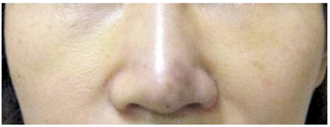
Fig. 1 After an autologous fat injection in the left periocular area, the skin color of the patient's nose changed to purple.
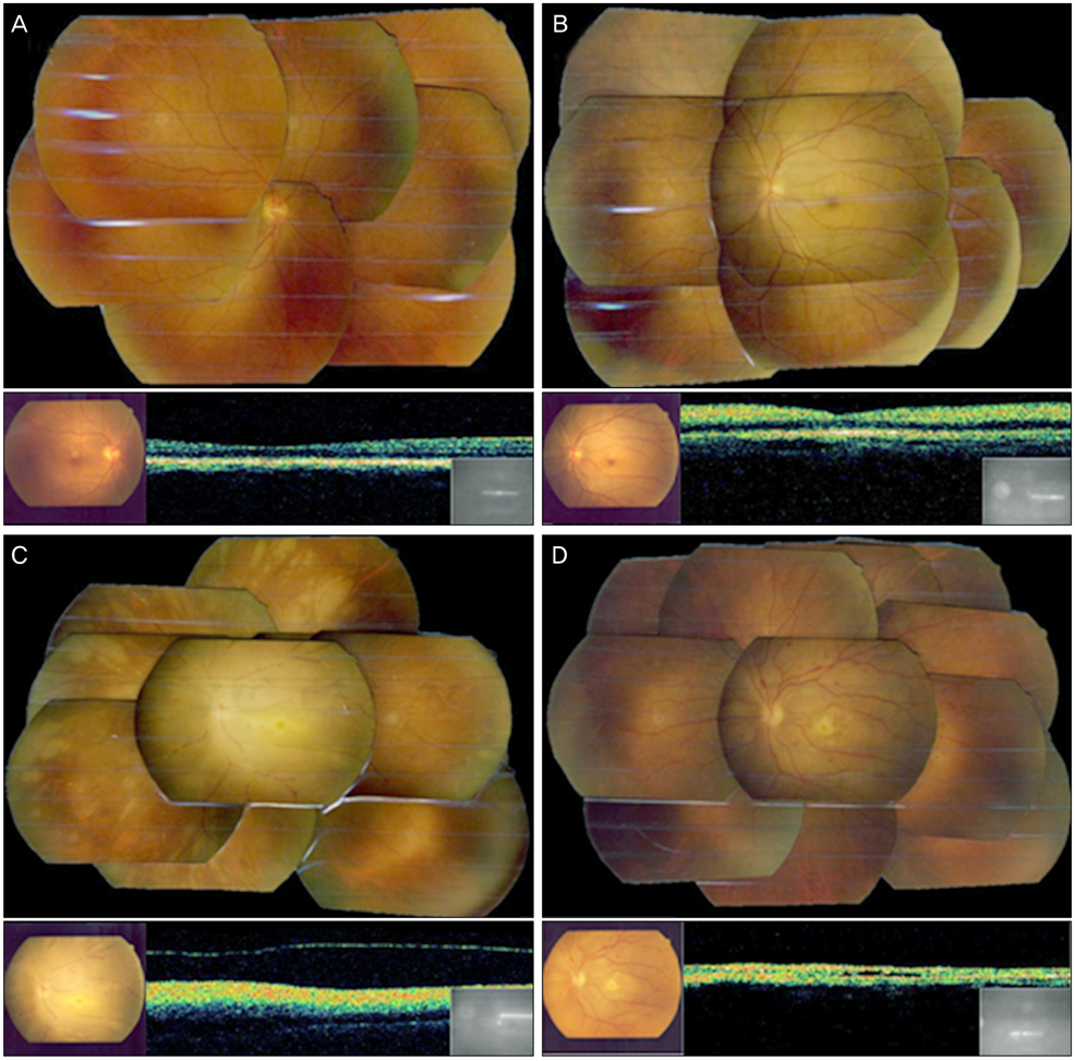
Fig. 2 (A) The fundus photo of the right eye shows no abnormal findings. (B) At 3 hours after the autologous fat injection, a photo of the fundus of the left eye shows a cherry red spot with visible emboli in several retinal arteries. (C) At 24 hours after the injection, a photo of the fundus of the left eye shows marked retinal edema, disc swelling and multiple fat emboli. (D) At 2 months after the injection, a photo of the fundus of the left eye shows optic disc atrophy, multiple retinal hemorrhages and a fibrous change on its posterior pole.
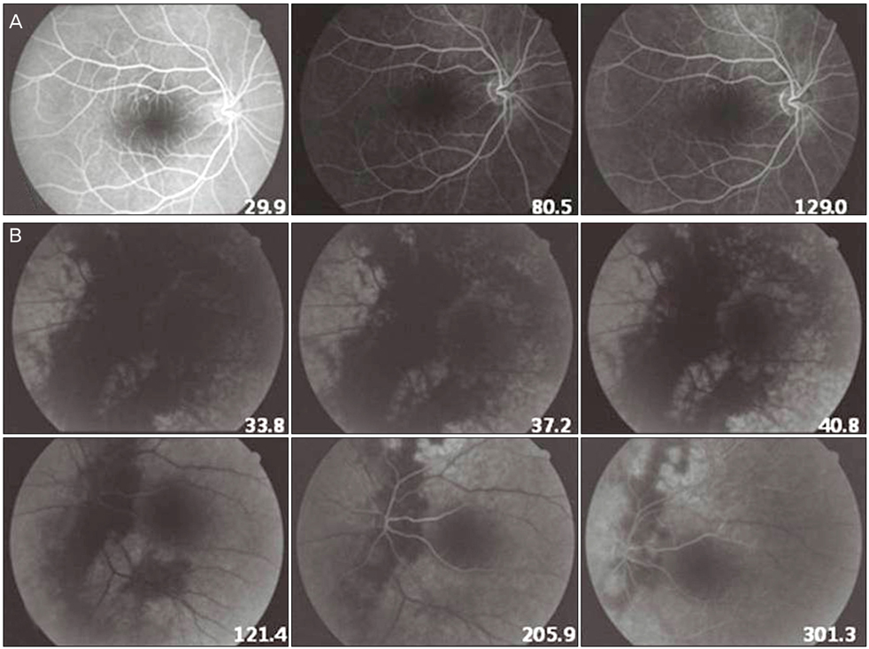
Fig. 3 (A) Fluorescein angiography revealed no abnormalities in the right eye, (B) but in the left eye it showed markedly prolonged choroidal filling around the optic disc and delays in both retinal arterial filling and retinal arteriovenous transit time.

Fig. 4 (A) The diffusion weighted brain magnetic resonance imaging showed an ill-defined hyperintense lesion at the left insular cortex and (B) decreased signal intensity at the same lesion on the afferent diffusion coefficient map. These results indicate recent brain infarction.
Cited by 2 articles
-
A Case of Visual Loss and Ophthalmoplegia Following Injection of Hyaluronic Acid into the Glabella
Dong Won Paik, In Bum Jang, Jae Suk Kim, Joo Hwa Lee, Jin Choi
J Korean Ophthalmol Soc. 2013;54(6):971-976. doi: 10.3341/jkos.2013.54.6.971.Multiple Cerebral Infarctions with Neurological Symptoms and Ophthalmic Artery Occlusion after Filler Injection
Won Sup Lee, Won Tae Yoon, Youn Joo Choi, Sung Pyo Park
J Korean Ophthalmol Soc. 2015;56(2):285-290. doi: 10.3341/jkos.2015.56.2.285.
Reference
-
1. Ellis PP. Occlusion of the central retinal artery after retrobulbar corticosteroid injection. Am J Ophthalmol. 1978. 85:352–356.2. Whiteman DW, Rosen DA, Pinkerton RM. Retinal and choroidal microvascular embolism after intranasal corticosteroid injection. Am J Ophthalmol. 1980. 89:851–853.3. Dreizen NG, Framm L. Sudden unilateral visual loss after autologous fat injection into the glabellar area. Am J Ophthalmol. 1989. 107:85–87.4. Danesh-Meyer HV, Savino PJ, Sergott RC. Case reports and small case series: ocular and cerebral ischemia following facial injection of autologous fat. Arch Ophthalmol. 2001. 119:777–778.5. Egido JA, Arroyo R, Marcos A, Jimenez-Alfaro I. Middle cerebral artery embolism and unilateral visual loss after autologous fat injection into the glabellar area. Stroke. 1993. 24:615–616.6. Park SH, Sun HJ, Choi KS. Sudden unilateral visual loss after autologous fat injection into the nasolabial fold. Clin Ophthalmol. 2008. 2:679–683.7. Brown GC, Magargal LE, Sergott R. Acute obstruction of the retinal and choroidal circulations. Ophthalmology. 1986. 93:1373–1382.8. Lee YJ, Kim HJ, Choi KD, Choi HY. MRI restricted diffusion in optic nerve infarction after autologous fat transplantation. J Neuroophthalmol. 2010. 30:216–218.
- Full Text Links
-
- Actions
-
Cited
- CITED
-
- Close
- Share
-
- Similar articles
-
- A Case of Acute Stroke after Autologous Fat Injection
- Retinal and Choroidal Vaseular Occlusion Following Autologous Fat Injection into the Temple Area
- Acute Cerebral Infarction and Visual Loss after Autologous Fat Injection into the Face
- Nasolacrimal Duct Obstruction Following Midfacial Autologous Fat Injection
- Correction of Sunken Eyelid with Unfavorable Fold Using Autologous Fat Injection
