Immunohistochemical study of osteopontin in boar testis
- Affiliations
-
- 1Department of Veterinary Medicine and Applied Radiological Science Research Institute, Cheju National University, Jeju 690-756, Korea. shint@cheju.ac.kr
- KMID: 1089659
- DOI: http://doi.org/10.4142/jvs.2007.8.2.107
Abstract
- The expression of osteopontin (OPN) in boar testis was studied. Western blot analysis detected 66- and 32-kDa OPN immunopositive bands in the testes of adult boars. In postnatal piglets, the 66-kDa OPN band was detected in the testes, but not the 32-kDa band. In the newborn testis, OPN immunostaining was seen in gonocytes and in some supporting cells in the seminiferous tubules, as well as in interstitial Leydig cells. In the adult boar testis, OPN immunoreactivity was detected in seminiferous tubules with varying intensities. Intense OPN immunostaining was seen in the residual bodies and acrosomes in the spermatids while, occasionally, OPN immunostaining was seen in spermatogonia and various stage of spermatocytes but in few Sertoli cells in the seminiferous tubules. In addition, Leydig cells in adult boars were weakly immunostained with OPN. These findings suggest that OPN is detected in the majority of germ cells and is involved in spermatogenesis in boar testis.
Keyword
MeSH Terms
Figure
-
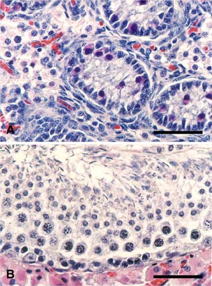
Fig. 1 Histology of the testes of postnatal (A) and adult (B) boars. (A) In the postnatal boar, sex cords and interstitial cells were seen. Undifferentiated supporting cells are positioned along the edges of the cords, while gonocytes, the precursors of spermatogonia, are located in the interior of the cords. (B) In the adult boar, there were interstitial cells and seminiferous tubules containing primary spermatocytes, spermatids, and Sertoli cells. H&E stain. bar = 50 µm.
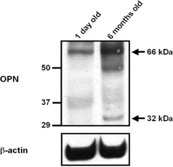
Fig. 2 A representative photo of a Western blot analysis of osteopontin (OPN) and beta-actin in the testes of postnatal and adult boars. The molecular sizes of standard proteins are shown on the left. Major and minor OPN bands were observed at the expected sizes of 66 and 32-kDa, respectively.
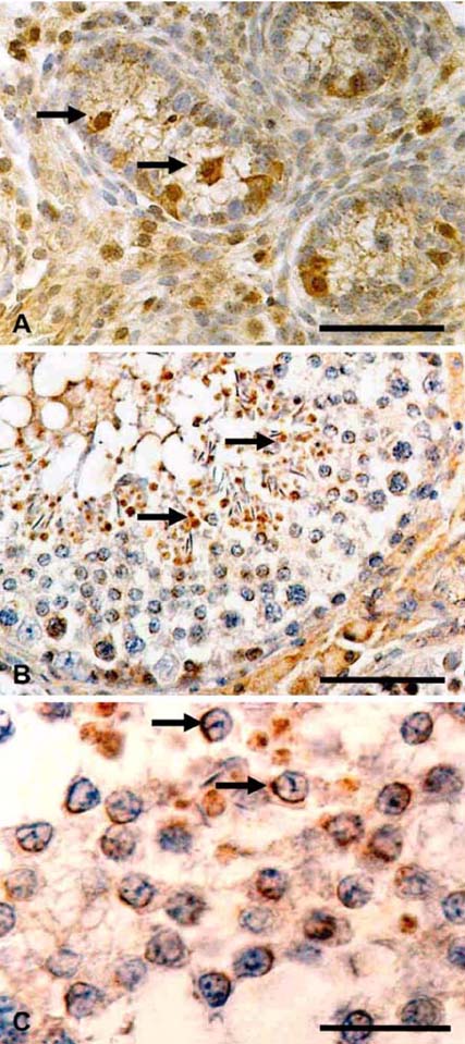
Fig. 3 Immunohistochemical analysis of osteopontin in the testes of postnatal (1 day old) (A) and adult (B, C) boars. (A) In the postnatal testis, osteopontin diffusely immunostained the testis, including the seminiferous tubules and interstitial cells. Gonocytes (arrows) in the undifferentiated tubules are typically immunopositive for OPN. (B) In the adult boar, intense osteopontin immunostaining was seen in the residual bodies (arrows) and acrosomes of spermatids in the seminiferous tubules, while OPN immunostaining was seen in spermatogonia, and various stage of spermatocytes as well as in some Leydig cells. (C) OPN-positive acrosomes (arrows) in spermatids are well visualized in an adjacent tubule. A-B; Counterstained with hematoxylin. bar = 50 µm (A and B), 20 µm (C).
Reference
-
1. Cancel AM, Chapman DA, Killian GJ. Osteopontin localization in the Holstein bull reproductive tract. Biol Reprod. 1999. 60:454–460.2. Denhardt DT, Guo X. Osteopontin: a protein with diverse functions. FASEB J. 1993. 7:1475–1482.
Article3. Goncalves RF, Wolinetz CD, Killian GJ. Influence of arginine-glycine-aspartic acid (RGD), integrins (αV and α5) and osteopontin on bovine sperm-egg binding, and fertilization in vitro. Theriogenology. 2007. 67:468–474.
Article4. Lin C, Tholen E, Jennen D, Ponsuksili S, Schellander K, Wimmers K. Evidence for effects of testis and epididymis expressed genes on sperm quality and boar fertility traits. Reprod Domest Anim. 2006. 41:538–543.
Article5. Luedtke CC, McKee MD, Cyr DG, Gregory M, Kaartinen MT, Mui J, Hermo L. Osteopontin expression and regulation in the testis, efferent ducts, and epididymis of rats during postnatal development through to adulthood. Biol Reprod. 2002. 66:1437–1448.
Article6. Moon C, Shin T. Increased expression of osteopontin in the spinal cords of Lewis rats with experimental autoimmune neuritis. J Vet Sci. 2004. 5:289–293.
Article7. Nagatomo T, Ohga S, Takada H, Nomura A, Hikino S, Imura M, Ohshima K, Hara T. Microarray analysis of human milk cells: persistent high expression of osteopontin during the lactation period. Clin Exp Immunol. 2004. 138:47–53.
Article8. Oldberg A, Franzen A, Heinegard D. Cloning and sequence analysis of rat bone sialoprotein (osteopontin) cDNA reveals an Arg-Gly-Asp cell-binding sequence. Proc Natl Acad Sci USA. 1986. 83:8819–8823.
Article9. Rodríguez CM, Day JR, Killian GJ. Osteopontin gene expression in the Holstein bull reproductive tract. J Androl. 2000. 21:414–420.10. Shin T, Ahn M, Kim H, Moon C, Kang TY, Lee JM, Sim KB, Hyun JW. Temporal expression of osteopontin and CD44 in rat brains with experimental cryolesions. Brain Res. 2005. 1041:95–101.
Article11. Siiteri JE, Ensrud KM, Moore A, Hamilton DW. Identification of osteopontin (OPN) mRNA and protein in the rat testis and epididymis, and on sperm. Mol Reprod Dev. 1995. 40:16–28.
Article12. Swierstra EE. Cytology and duration of the cycle of the seminiferous epithelium of the boar; duration of spermatozoan transit through the epididymis. Anat Rec. 1968. 161:171–185.
Article13. Wai PY, Kuo PC. The role of osteopontin in tumor metastasis. J Surg Res. 2004. 121:228–241.
Article14. Weber GF, Cantor H. The immunology of Eta-1/osteopontin. Cytokine Growth Factor Rev. 1996. 7:241–248.
Article15. Wilson MJ, Liaw L, Koopman P. Osteopontin and related SIBLING glycoprotein genes are expressed by Sertoli cells during mouse testis development. Dev Dyn. 2005. 233:1488–1495.
Article16. Xie Y, Sakatsume M, Nishi S, Narita I, Arakawa M, Gejyo F. Expression, roles, receptors, and regulation of osteopontin in the kidney. Kidney Int. 2001. 60:1645–1657.
Article
- Full Text Links
-
- Actions
-
Cited
- CITED
-
- Close
- Share
-
- Similar articles
-
- The Immunohistochemical Patterns of Calcification-related Molecules in the Epidermis and Dermis of the Zebrafish (Danio rerio)
- Identification of African swine fever virus genomic DNAs in wild boar habitats within outbreak regions in South Korea
- Osteopontin expression on benign and malignant ovarian tumors
- Expression of Osteopontin in Osteoclast
- Effects of Osteopontin on Normal and Malignant Ovarian Epithelial Cell
