Radiographic and computed tomographic evaluation of experimentally induced lung aspiration sites in dogs
- Affiliations
-
- 1Department of Veterinary Diagnostic Imaging, College of Veterinary Medicine, Konkuk University, Seoul 143-701, Korea. eomkd@konkuk.ac.kr
- 2Department of Veterinary Surgery, College of Veterinary Medicine, Kyungpook National University, Daegu 702-701, Korea.
- KMID: 1089490
- DOI: http://doi.org/10.4142/jvs.2006.7.4.397
Abstract
- This study was performed to radiographically examine the prevalence of aspiration sites and to evaluate their atomical correlation with the bronchial pattens. Ten healthy beagle dogs were repeatedly radiographed, at weekly intervals, in the left and right lateral, ventrodorsal (VD) and dorsoventral (DV) positions. Three mililiters of iohexol distilled with same volume of saline was infused into the tracheal inlet. Which lung lobe was aspirated was decided upon by the presence of a significant alveolar pattern due to the contrast medium. Alveolar patterns were identified at the left (100%) and right cranial lung lobes (77%) with the dogs in dependant lateral recumbency, at the right caudal lung lobe (71%) with the dogs in VD recumbency and at the right middle lung lobe (59%) with the dogs in DV recumbency, respectively. The anatomical correlation was evaluated by performing computed tomography. The right principal bronchus (165.8 +/- 1.6 degrees) was more straightly bifurcated than was the left principal bronchus (142.7 +/- 1.8 degrees, p < 0.01). In VD position, the right side lung had a greater opertunity to become aspirated. The ventrally positioned right middle lobar bronchial origin was more easily to be aspirated the other laterally positioned ones. We think that these anatomical characteristics can be one of the causes for aspiration pneumonia to occur more frequently in the right side lung.
Keyword
MeSH Terms
Figure
-
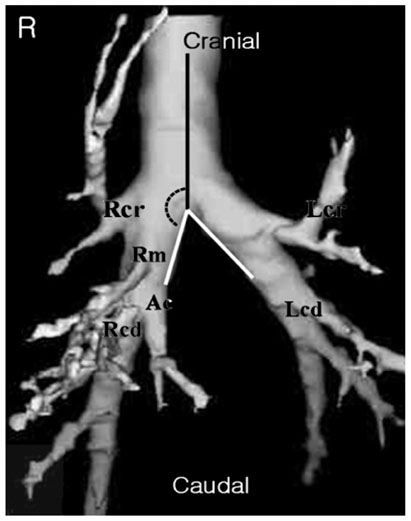
Fig. 1 Ventral aspect view of the three-dimensional reconstruction CT images. The right cranial (Rcr), middle (Rm), caudal (Rcd) and accessory (Ac) lobar bronchi in the right side and the cranial (Lcr) and caudal (Lcd) lobar bronchi in the left side are seen. The middle lobar bronchus originates from the ventral side of the right principal bronchus. The angle (black dotted curved line) was measured between the principal bronchial (white line) and the tracheal (black line) extension.
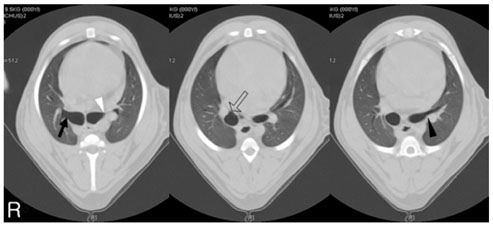
Fig. 2 CT images of the canine thorax in ventrodorsal recumbency. The right cranial (black arrow), middle (open arrow), the beginning (white arrow head) and the full sliced diameter (black arrow head) of the left cranial bronchial opening are visualized.
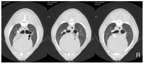
Fig. 3 CT images of the canine thorax in dorsoventral recumbency. The right cranial (black arrow), middle (open arrow) and left cranial (white arrow head) bronchial openings are seen.
Reference
-
1. Burk RL, Ackerman N. Burk RL, Ackerman N, editors. The thorax. Small Animal Radiology and Ultrasonography. 1996. 2nd ed. Philadelphia: Saunders;25–248.
Article2. Farrow CS. Farrow CS, editor. Pneumonia. Veterinary Diagnostic Imaging the Dog and Cat. 2003. St. Louis: Mosby;407–418.
Article3. Ford RB. Tams TR, editor. Normal canine and feline trachea and bronchi. Small Animal Endoscopy. 1990. St. Louis: Mosby;297–326.4. Hawkins EC. Bonagura JD, editor. Aspiration pneumonia. Current Veterinary Therapy XIV. 1995. Philadelphia: Saunders;915–919.5. Marik PE. Aspiration pneumonitis and aspiration pneumonia. N Engl J Med. 2001. 344:665–671.
Article6. Moore DE, Carroll FE, Dutt PL, Reed GW, Holburn GE. Comparison of nonionic and ionic contrast agents in the rabbit lung. Invest Radiol. 1991. 26:134–142.
Article7. Nakakuki S. The bronchial tree and lobular division of the dog lung. J Vet Med Sci. 1994. 56:455–458.
Article8. Nelson OL, Sellon RK. Ettinger SJ, Feldman EC, editors. Pulmonary parenchymal disease. Textbook of Veterinary Internal Medicine. 2005. 6th ed. St. Louis: Eslvier;1239–1266.9. Nelson RW, Couto CG. Nelson RW, Couto CG, editors. Disorders of the pulmonary parenchyma. Small Animal Internal Medicine. 2003. 3rd ed. St. Louis: Mosby;299–314.
- Full Text Links
-
- Actions
-
Cited
- CITED
-
- Close
- Share
-
- Similar articles
-
- Effects of Extracorporeal Shock Wave Lithortripsy Experimentally Induced Cholelithiasis and Organs in the Dog
- Virtual otoscopy for evaluating the inner ear with a fluid-filled tympanic cavity in dogs
- Computed tomographic bronchioarterial ratio for brachycephalic dogs without pulmonary disease
- Radiographic diagnosis of diaphragmatic hernia: review of 60 cases in dogs and cats
- A Radiologic Overview of Aspiration-Induced Lung Disease in Adults
