J Vet Sci.
2009 Mar;10(1):77-80. 10.4142/jvs.2009.10.1.77.
Ultrasonographic examination of the carpal canal in dogs
- Affiliations
-
- 1Department of Anatomy, Faculty of Veterinary Medicine, University of Adnan Menderes, PK: 17, 09016, Isikli-Aydin, Turkey. terkut@yahoo.com
- 2Department of Radiology, Faculty of Medicine, University of Adnan Menderes, PK: 17, 09016, Isikli-Aydin, Turkey.
- KMID: 1089350
- DOI: http://doi.org/10.4142/jvs.2009.10.1.77
Abstract
- The aim of this study was to determine the course of the median nerve and its adjacent structures in the carpal canals of 8 healthy dogs by using high-frequency transducers. Before performing ultrasonography, the transverse and posteroanterior diameters as well as the perimeter of the carpus were measured at just proximal to the side of the carpal pad. The anatomical structures were then determined at two levels of the carpal canal, which were named the proximal and distal levels, on the transverse sonograms. The cross-sectional areas, perimeters and the transverse and posteroanterior diameters of the median nerve were measured at these levels. Although all the measurements were larger at the proximal level, significant differences between the proximal and distal levels were determined for the cross-sectional area, the perimeter and the transverse diameter of the median nerve. On the transverse sonogram, the deep digital flexor tendon was seen in almost the center of the carpal canal like a comma shape and also it had a small concavity on the caudal side. The superficial digital flexor tendon was seen as an ovoid shape on the transverse sonograms and it was located nearly at the posterior side of the carpal canal. Both tendons were seen as intermediate-grade echogenic structures. The median artery was located inside of the concavity of the deep digital flexor tendon. Also, the median nerve was seen at the posteromedial side of the median artery. As a result of this study, the cross-sectional areas of the median nerve ranged between 1.01-2.68 mm2 at the proximal level and between 0.93-1.91 mm2 at the distal level.
Keyword
Figure
-
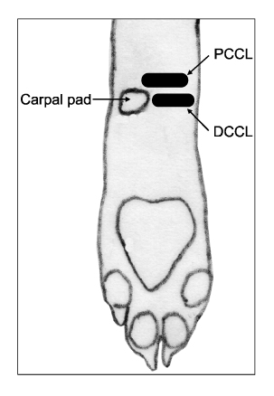
Fig. 1 Schematic presentation of the probe placement for the proximal (PCCL) and distal (DCCL) carpal canal levels in the left forelimb.
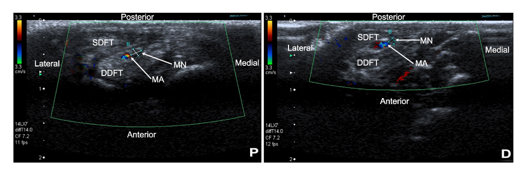
Fig. 2 Transverse ultrasonographic scans of the carpal canal at the proximal (P) and the distal (D) levels of the carpal canal. Color Doppler imaging is helpful to easily localize the median nerve. Note that the size of the median nerve is larger at the proximal part of the carpal canal as compared to the distal part. The transverse diameter (A) and the posteroanterior diameter (B) of the median nerve, the median nerve (MN), the median artery (MA), the deep digital flexor (DDFT) tendons and the superficial digital flexor tendons (SDFT).
Reference
-
1. Alexander K, Dobson H. Ultrasonography of peripheral nerves in the normal adult horse. Vet Radiol Ultrasound. 2003. 4:456–464.
Article2. Beekman R, Visser LH. High-resolution sonography of the peripheral nervous system - a review of the literature. Eur J Neurol. 2004. 11:305–314.
Article3. Benigni L, Corr SA, Lamb CR. Ultrasonographic assessment of the canine sciatic nerve. Vet Radiol Ultrasound. 2007. 48:428–433.
Article4. Bleecker ML, Bolhman M, Moreland R, Tipton A. Carpal tunnel syndrome: role of carpal canal size. Neurology. 1985. 35:1599–1604.
Article5. Buchberger W, Schon G, Strasser K, Jungwirth W. High-resolution ultrasonography of the carpal tunnel. J Ultrasound Med. 1991. 10:531–537.
Article6. El Miedany YM, Aty SA, Ashour S. Ultrasonography versus nerve conduction study in patients with carpal tunnel syndrome: substantive or complementary tests? Rheumatology. 2004. 43:887–895.
Article7. Evans HE, Christensen GC. Miller's Anatomy of the Dog. 1979. Philadelphia: Saunders;248.8. Hudson JA, Steiss JE, Braund KG, Toivio-Kinnucan M. Ultrasonography of peripheral nerves during wallerian degeneration and regeneration following transection. Vet Radiol Ultrasound. 1996. 37:302–312.
Article9. Hudson JA, Finn-Bodner ST, Steiss JE. Neurosonography. Vet Clin North Am Small Anim Pract. 1998. 28:943–972.
Article10. Kramer M, Gerwing M, Hach V, Schimke E. Sonography of the musculoskeletal system in dogs and cats. Vet Radiol Ultrasound. 1997. 38:139–149.
Article11. Lamb CR, Wong K. Ultrasonographic anatomy of the canine elbow. Vet Radiol Ultrasound. 2005. 46:319–325.
Article12. Lee CH, Kim TK, Yoon ES, Dhong ES. Correlation of high-resolution ultrasonographic findings with the clinical symptoms and electrodiagnostic data in carpal tunnel syndrome. Ann Plast Surg. 2005. 54:20–23.
Article13. Lee HC, Choi HJ, Choi MC, Yoon JH. Ultrasonographic measurement of optic nerve sheath diameter in normal dogs. J Vet Sci. 2003. 4:265–268.
Article14. Mackay-Smith MP, Cushing LS, Leslie JA. "Carpal canal" syndrome in horses. J Am Vet Med Assoc. 1972. 160:993–997.15. Martinoli C, Bianchi S, Gandolfo N, Valle M, Simonetti S, Derchi LE. US of nerve entrapments in osteofibrous tunnels of the upper and lower limbs. Radiographics. 2000. 20:S199–S213.
Article16. Mazurek MT, Shin AY. Upper extremity peripheral nerve anatomy: current concepts and applications. Clin Orthop Relat Res. 2001. 383:7–20.17. Preston DC. Distal median neuropathies. Neurol Clin. 1999. 17:407–424.
Article18. Rose S, Long C, Knipe M, Hornof B. Ultrasonographic evaluation of brachial plexus tumors in five dogs. Vet Radiol Ultrasound. 2005. 46:514–517.
Article19. Squire KR, Adams SB, Widmer WR, Coatney RW, Habig C. Arthroscopic removal of a palmar radial osteochondroma causing carpal canal syndrome in a horse. J Am Vet Med Assoc. 1992. 201:1216–1218.20. Turan E, Bolukbasi O. Evaluation of possible carpal tunnel syndrome in dogs. Vet Rec. 2004. 155:122–124.
Article21. Turan E, Erden H. Computed tomography and morphometry of the carpal canal in the dog. Ann Anat. 2003. 185:173–178.
Article22. Walker FO, Cartwright MS, Wiesler ER, Caress J. Ultrasound of nerve and muscle. Clin Neurophysiol. 2004. 115:495–507.
Article23. Winn FJ Jr, Habes DJ. Carpal tunnel area as a risk factor for carpal tunnel syndrome. Muscle Nerve. 1990. 13:254–258.
Article24. Yamanaka K. An experimental study on compression neuropathy-the effect of neurolysis on the blood-nerve barrier. Nippon Seikeigeka Gakkai Zasshi. 1992. 66:714–727.25. Yesildag A, Kutluhan S, Sengul N, Koyuncuoglu HR, Oyar O, Guler K, Gulsoy UK. The role of ultrasonographic measurements of the median nerve in the diagnosis of carpal tunnel syndrome. Clin Radiol. 2004. 59:910–915.
Article26. Ziswiler HR, Reichenbach S, Vögelin E, Bachmann LM, Villiger PM, Jüni P. Diagnostic value of sonography in patients with suspected carpal tunnel syndrome: a prospective study. Arthritis Rheum. 2005. 52:304–311.
Article
- Full Text Links
-
- Actions
-
Cited
- CITED
-
- Close
- Share
-
- Similar articles
-
- Ultrasonographic Study of Median Nerve after Carpal Tunnel Release
- Ultrasonographic Findings of Median Nerve Change according to the Wrist Position
- The Correlation Between Electrodiagnostic Results and Ultrasonographic Findings in the Severity of Carpal Tunnel Syndrome in Females
- Subclinical Ulnar Neuropathy in Carpal Tunnel Syndrome
- Carpal Tunnel Syndrome
