Distribution and ontogeny of gastrin- and serotonin-immunoreactive cells in the proventriculus of developing chick, Gallus gallus domestica
- Affiliations
-
- 1Histology and Pathology Laboratory, Mediterranean Fisheries Research Production and Education Institute, PK 190, Antalya, 07001, Turkey. aksoy@akdenizsuurunleri.gov.tr
- 2Department of Biology, Suleyman Demirel University, Isparta, 32260, Turkey.
- KMID: 1089340
- DOI: http://doi.org/10.4142/jvs.2009.10.1.9
Abstract
- The ontogeny and distribution of gastrin- and serotonin-immunoreactive (IR) cell in the proventriculus of chicks (Gallus gallus domestica, n = 60) in different growth periods was examined immunohistochemically using antisera specific to gastrin and serotonin. Gastrin and serotonin-IR cells were detected in chick proventriculus. Gastrin-IR cells were first evident after 12 days of incubation in lamina epithelialis and compound glands, while serotonin-IR cells were observed only in compound glands at that same time. Gastrin-IR and serotonin-IR cells increased in frequency on incubation day 14 and 16, respectively. Towards the end of incubation, gastrin- and serotonin-IR cell numbers decreased. In adult chicken, both IR cells were present but not lower numbers. The observations demonstrate the presence of gastrin- and serotonin-IR cells in the proventriculus of developing chicks in temporally changing frequencies.
Keyword
MeSH Terms
Figure
-
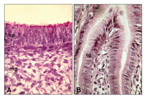
Fig. 1 The superficial epithelium of the proventriculus. The superficial epithelium of proventriculus, which was lined by a pseudostratified columnar-like epithelium on day 9 of incubation (A), varied to simple columnar epithelium by day 14 of incubation (B). mct: mesenchymal connective tissue, ep: epithelium, lct: loose connective tissue. Masson trichrome stain. ×1,200.
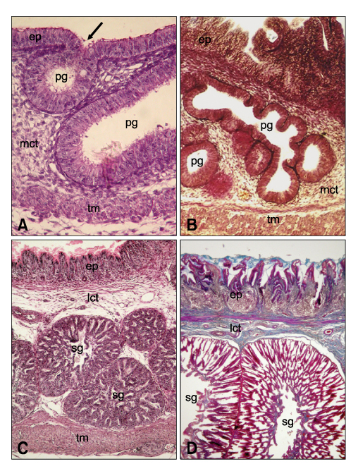
Fig. 2 Histological structure of proventriculus wall in different development stages. Primordial glands that were initially evident as an epithelial invagination on day 9 of incubation (A), appearred branching by day 11 of incubation (B). With development, the enlargement in the size of the stomach glands and the increase in thickness of the proventriculus wall were seen clearly in chicks in hatching time (C) and in adults (D). ep: epithelium, mct: mesenchymal connective tissue, lct: loose connective tissue, pg: primordial gland, sg, stomach gland, tm: tunica muscularis. Arrow indicates epithelial invagination constituting stomach glands in the next development stages. Masson trichrome stain. A, ×400, B, ×220, C, ×150, D, ×60.
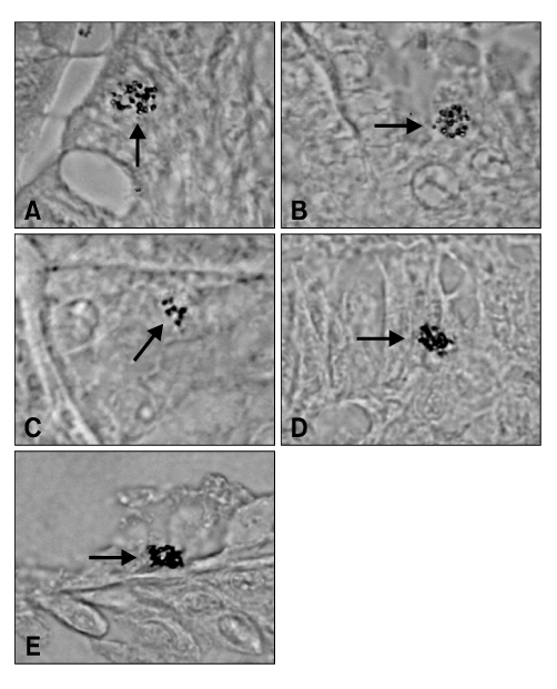
Fig. 3 Localization of gastrin- and serotonin-immunoreactive (IR) cells. Gastrin-IR cells were evident in the proventriculus surface epithelium (A) and compound gland (B) on day 12 of incubation. (C) Serotonin-IR cells were evident in the compound gland beginning from day 12 of incubation. In adults, gastrin- (D) and serotonin-IR cells (E) were present in epithelium and compound glands, respectively. Arrows indicate gastrin- or serotonin-IR cells. Avidin-biotin-peroxidase method. A, B and D, ×1,300, C, ×800, E, ×1,500.
Reference
-
1. Alison BC. The distribution and ontogeny of gastrin/CCK-, somatostatin- and neurotensin-immunoreactive cells in the gastrointestinal tract of the chicken. Histol Histopathol. 1989. 4:55–62.2. Alison BC. The ontogeny and distribution of glucagon- and pancreatic polypeptide-immunoreactive cells in the gastrointestinal tract of the chicken. Anat Embryol. 1990. 182:605–610.
Article3. Beorlegui C, Martínez A, Sesma P. Endocrine cells and nerves in the pyloric ceca and the intestine of Oncorhynchus mykiss (Teleostei): An immunocytochemical study. Gen Comp Endocrinol. 1992. 86:483–495.
Article4. Castaldo L, Lucini C. An immunohistochemical study on the endocrine cells in the gastrointestinal tract of domestic duck. Eur J Basic Appl Histochem. 1991. 35:131–143.5. Castaldo L, Lucini C. Ontogenesis of some endocrine cells in the duck gastrointestinal tract. Eur J Histochem. 1994. 38:319–326.6. Catroxo MHB, Lima MAI, Petrella S. Ultrastructure of endocrine cells of the stomach (proventriculus and gizzard) of the red-capped cardinal (Paroaria g. gularis, Linnaeus, 1766). Rev Chil Anat. 2001. 19:239–244.7. D'este L, Biancone S, Renda T. Ontogenesis of 5-hydroxytryptamine-like immunoreactive cells and their relationship with bombesin in chicken proventriculus. Basic Appl Histochem. 1986. 30:109–117.8. Dimaline R, Lee CM. Chicken Gastrin: A member of the gastrin/CCK family with novel structure-activity relationships. Am J Physiol. 1990. 259:G882–G888.
Article9. Dockray GJ. Gastrin and gastric epithelial physiology. J Physiol. 1999. 518:315–324.10. Gulmez N, Nazli M, Aslan S, Liman N. Immunolocalisation of serotonin, gastrin, somatostatin and glucagon in enteroendocrine cells of the goose (Anser anser). Acta Vet Hung. 2003. 51:439–449.
Article11. Hsu SM, Raine L, Fanger H. Use of avidin-biotin-peroxidase complex (ABC) in immunoperoxidase techniques: a comparison between ABC and unlabeled antibody (PAP) procedures. J Histochem Cytochem. 1981. 29:577–580.
Article12. Larsson LI. Developmental biology of gastrin and somatostatin cells in the antropyloric mucosa of the stomach. Microsc Res Tech. 2000. 48:272–281.
Article13. Lee HS, Ku SK. An immunohistochemical study of endocrine cells in the alimentary tract of the grass lizard, Takydromus wolteri Fischer (Laceridae). Acta Histochem. 2004. 106:171–178.
Article14. Lee JH, Ku SK, Lee HS, Kitagawa H. An immunohistochemical study of endocrine cells in the pancreas of the Red-bellied frog (Bombina orientalis). Eur J Histochem. 2003. 47:165–172.
Article15. Luna LG. Carson FL, editor. Masson trichrome stain. Histotechnology: A Self Instructional Text. 1997. Chicago: American Society for Clinical Pathology Press;134.16. Martínez A, López J, Barrenechea MA, Sesma P. Immunocytochemical and ultrastructural characterization of endocrine cells in chicken proventriculus. Cell Tissue Res. 1991. 263:541–548.
Article17. Martínez A, López J, Sesma P. Development of the diffuse endocrine system in the chicken proventriculus. Cell Tissue Res. 1993. 271:107–113.
Article18. Martínez A, López J, Sesma P. The nervous system of the chicken proventriculus: an immunocytochemical and ultrastructural study. Histochem J. 2000. 32:63–70.19. Mensah-Brown EPK, Lawrence PA. Neurotransmitters regulating acid secretion in the proventriculus of the houbara bustard (Chlamydotis undulata): A morphological viewpoint. J Morphol. 2001. 248:175–184.
Article20. Neglia S, Arcamone N, Esposito V, Gargiulo G, De Girolamo P. Presence and distribution of ghrelin-immunopositive cells in the chicken gastrointestinal tract. Acta Histochem. 2005. 107:3–9.
Article21. Okamoto T, Fujii S. An electron microscopic study on endocrine cells in the pyloric region of the duck. Nippon Juigaku Zasshi. 1980. 42:169–176.
Article22. Peroutka SJ. 5-Hydroxytryptamine receptor subtypes. Annu Rev Neurosci. 1988. 11:45–60.
Article23. Rawdon BB. Gastrointestinal hormones in birds: morphological, chemical, and developmental aspects. J Exp Zool. 1984. 232:659–670.
Article24. Rawdon BB, Andrew A. An immunocytochemical survey of endocrine cells in the gastrointestinal tract of chicks at hatching. Cell Tissue Res. 1981. 220:279–292.
Article25. Rawdon BB, Andrew A. Distribution of serotonin-immunoreactive gut endocrine cells in chicks at hatching. examination of possible co-localisation with peptides reveals unexpected cross-reactivity of substance P antiserum with serotonin. Histochemistry. 1994. 102:93–100.
Article26. Rawdon BB, Andrew A. Gut endocrine cells in birds: an overview, with particular reference to the chemistry of gut peptides and the distribution, ontogeny, embryonic origin and differentiation of the endocrine cells. Prog Histochem Cytochem. 1999. 34:3–82.
Article27. Richardson BP, Engel G. The pharmacology and function of 5-HT3 receptors. Trends Neurosci. 1986. 9:424–428.28. Salvi E, Vaccaro R, Renda TG. An immunohistochemical study of the ontogeny of the neuroendocrine system in the chicken oesophagus. Anat Embryol. 1998. 197:283–291.
Article29. Timurkaan S, Karan M, Aydin A. Immunohistochemical study of the distribution of serotonin in the gastrointestinal tract of the porcupines (Hystrix cristata). Rev Med Vet. 2005. 156:533–536.30. Yamaguchi S, Yamada J, Kitamura N, Yamashita T. Ontogeny of the endocrine cells in the quail proventriculus. Z Mikrosk Anat Forsch. 1986. 100:981–989.31. Yaman M, Tarakçi BG, Bayrakdar A, Yaman I. Immunohistochemical study of the endocrine cells in the oesophagus of the ostrich (Struthio camelus). Rev Med Vet. 2008. 159:63–67.32. Yamanaka Y, Yamada J, Kitamura N, Yamashita T. An immunohistochemical study on the distribution of endocrine cells in the chicken gastrointestinal tract. Z Mikrosk Anat Forsch. 1989. 103:437–446.
- Full Text Links
-
- Actions
-
Cited
- CITED
-
- Close
- Share
-
- Similar articles
-
- A new cestode Raillietina (Skrjabinia) doggaddaensis n. sp. from Gallus gallus domesticus (L.) from India
- Immunohistochemical Studies on the Serotonin, Somatostatin and Gastrin-Positive Cells in the Gastric Adenocarcinoma
- Morphological Changes of Endolymphatic Sac after Gentamicin Injection to Chick Embryo
- Regional Distribution and Relative Frequency of Gastrointestinal Endocrine Cells in Large Intestines of C57BL/6 Mice
- Immunocytochemical study on the somatostatin,serotonin and gastrin cells in the gastrointestinal tract of the percida
