Korean J Radiol.
2009 Apr;10(2):190-193. 10.3348/kjr.2009.10.2.190.
Prenatal Diagnosis of Transient Abnormal Myelopoiesis in a Down Syndrome Fetus
- Affiliations
-
- 1Department of Obstetric and Gynecology, Chung-Ang University School of Medicine, Seoul, Korea. les740822@yahoo.co.kr
- KMID: 1088731
- DOI: http://doi.org/10.3348/kjr.2009.10.2.190
Abstract
- We report a case of transient abnormal myelopoiesis in a Down syndrome fetus diagnosed at 28(+3) weeks of gestation that rapidly progressed to intrauterine death 10 days later. Fetal hepatosplenomegaly with cerebral ventriculomegaly, although not specific, may be a suggestive finding of Down syndrome with transient abnormal myelopoiesis. Prompt fetal blood sampling for liver function test and chromosomal analysis are mandatory for early detection and management.
MeSH Terms
Figure
-
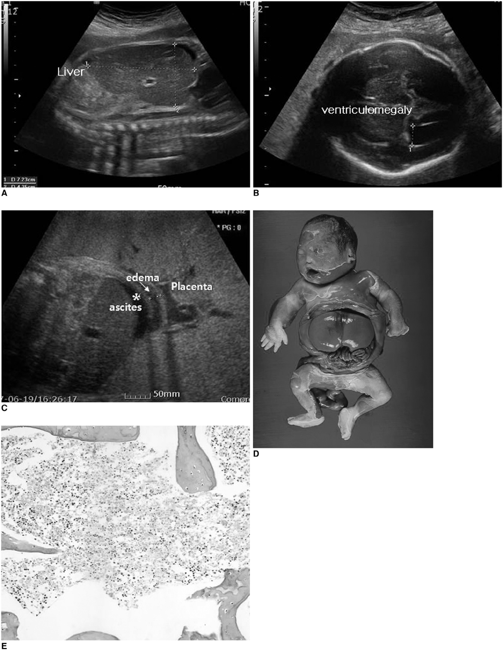
Fig. 1 Transient abnormal myelopoiesis in Down syndrome fetus. A. Longitudinal ultrasound scan of fetal abdomen shows hypoechoic liver occupying nearly entire enlarged abdomen. Liver length of 7.2 cm represents 90th percentile size. B. Fetal cerebral ventriculomegaly measuring 1.1 cm is evident. C. Oblique ultrasound scan of fetal abdomen. Skin edema (→) and ascites (*) are evident at 29+4 weeks' gestation. D. Postmortem picture of fetus with hepatomegaly. E. Myeloblastic cells infiltrating bone marrow (Hematoxylin & Eosin staining, ×40 magnification).
Reference
-
1. Hojo S, Tsukimori K, Kitade S, Nakanami N, Hikino S, Hara T, et al. Prenatal sonographic findings and hematological abnormalities in fetuses with transient abnormal myelopoiesis with Down syndrome. Prenat Diagn. 2007. 27:507–511.2. Smrcek JM, Baschat AA, Germer U, Gloeckner-Hofmann K, Gembruch U. Fetal hydrops and hepatosplenomegaly in the second half of pregnancy: a sign of myeloproliferative disorder in fetuses with trisomy 21. Ultrasound Obstet Gynecol. 2001. 17:403–409.3. Hamada H, Yamada N, Watanabe H, Okuno S, Fujiki Y, Kubo T. Hypoechoic hepatomegaly associated with transient abnormal myelopoiesis provides clues to trisomy 21 in the third-trimester fetus. Ultrasound Obstet Gynecol. 2001. 17:442–444.4. Kikuchi A, Tamura N, Ishii K, Takakuwa K, Matsunaga M, Sudo N, et al. Four cases of fetal hypoechoic hepatomegaly associated with trisomy 21 and transient abnormal myelopoiesis. Prenat Diagn. 2007. 27:665–669.5. Hadlock FP, Deter RL, Harrist RB, Park SK. Estimating fetal age: computer-assisted analysis of multiple fetal growth parameters. Radiology. 1984. 152:497–501.6. Vintzileos AM, Neckles S, Campbell WA, Andreoli JW Jr, Kaplan BM, Nochimson DJ. Fetal liver ultrasound measurements during normal pregnancy. Obstet Gynecol. 1985. 66:477–480.7. Enid GB, Diane DS. Embryo and fetal pathology: color atlas with ultrasound correlation. 2004. NewYork: Cambridge University Press;665.8. Fong CT, Brodeur GM. Down's syndrome and leukemia: epidemiology, genetics, cytogenetics and mechanisms of leukemogenesis. Cancer Genet Cytogenet. 1987. 28:55–76.9. Zipursky A, Brown EJ, Christensen H, Doyle J. Transient myeloproliferative disorder (transient leukemia) and hematologic manifestations of Down syndrome. Clin Lab Med. 1999. 19:157–167.10. Ogawa M, Hosoya N, Sato A, Tanaka T. Is the degree of fetal hepatosplenomegaly with transient abnormal myelopoiesis closely related to the postnatal severity of hematological abnormalities in Down syndrome? Ultrasound Obstet Gynecol. 2004. 24:83–85.11. Robertson M, De Jong G, Mansvelt E. Prenatal diagnosis of congenital leukemia in a fetus at 25 weeks' gestation with Down syndrome: case report and review of the literature. Ultrasound Obstet Gynecol. 2003. 21:486–489.12. Pares D, Chinen PA, Camano L, Moron AF, Torloni MR. Prediction of fetal anemia by Doppler of the middle cerebral artery and descending thoracic aorta. Arch Gynecol Obstet. 2008. 278:27–31.13. Scheier M, Hernandez-Adnrade E, Carmo A, Dezerega V, Nicolaides KH. Prediction of fetal anemia in rhesus disease by measurement of fetal middle cerebral artery peak systolic velocity. Ultrasound Obstet Gynecol. 2004. 23:432–436.14. Hartung J, Chaoui R, Wauer R, Bollmann R. Fetal hepatosplenomegaly: an isolated sonographic sign of trisomy 21 in a case of myeloproliferative disorder. Ultrasound Obstet Gynecol. 1998. 11:453–455.15. Baschat AA, Wagner T, Malisius R, Gembruch U. Prenatal diagnosis of a transient myeloproliferative disorder in trisomy 21. Prenat Diagn. 1998. 18:731–736.16. Vimercati A, Greco P, Gentile A, Ingravallo G, Loverro G, Selvaggi L. Fetal liver hyperechogenicity on sonography may be a serendipitous sign of a transient myeloproliferating disorder. Prenat Diagn. 2003. 23:44–47.17. Shipp TD, Benacerraf BR. Second trimester ultrasound screening for chromosomal abnormalities. Prenat Diagn. 2002. 22:296–307.18. Haydar TF. Advanced microscopic imaging methods to investigate cortical development and the etiology of mental retardation. Ment Retard Dev Disabil Res Rev. 2005. 11:303–316.19. Forestier F, Daffos F, Galacteros F, Bardakjian J, Rainaut M, Beuzard Y. Hematological values of 163 normal fetuses between 18 and 30 weeks of gestation. Pediatr Res. 1986. 20:342–346.20. Gozzo ML, Noia G, Barbaresi G, Colacicco L, Serraino MA, De Santis M, et al. Reference intervals for 18 clinical chemistry analytes in fetal plasma samples between 18 and 40 weeks of pregnancy. Clin Chem. 1998. 44:683–685.
- Full Text Links
-
- Actions
-
Cited
- CITED
-
- Close
- Share
-
- Similar articles
-
- Prenatal diagnosis of a fetus with Klinefelter's syndrome
- Diagnosis of fetal anomalies by sonography
- A Review of Prenatal Cytogenetic Analysis in 2942 Midtrimester Amniocentesis
- Prenatal Diagnosis of A Case of Fetus in Fetu in the Fetal Retroperitoneum
- Clinical Analysis of 739 Cases of Midtrimester Amniocentesis
