A Case of Epithelial Inclusion Cyst of Iris
- Affiliations
-
- 1Department of Ophthalmology, St. Mary's Hospital, College of Medicine, The Catholic University of Korea, Seoul, Korea. sara514@catholic.ac.kr
- KMID: 1084215
- DOI: http://doi.org/10.3341/kjo.2008.22.4.259
Abstract
- To report on an epithelial inclusion cyst of the iris that was successfully treated with needle aspiration and Ab externo laser photocoagulation. A 6-year-old boy was treated for a 6.0 mm fluid-filled cyst in the anterior chamber of the right eye. Thirteen months previously, he had undergone primary closure of a 6 mm full-thickness corneal laceration. The subsequent cyst was diagnosed as an epithelial inclusion cyst of the iris. His vision decreased to finger-count at 30 cm as the cyst grew over the pupil. We performed needle aspiration of the cyst and Ab externo laser photocoagulation of the cyst wall. The treated lesion was completely removed. The patient's visual acuity recovered to 20/40 without complications. There was no recurrence as determined by slit lamp examination up to 6 months after treatment. Needle aspiration and Ab externo laser photocoagulation can be used to effectively treat epithelial inclusion cysts of the iris.
MeSH Terms
Figure
-
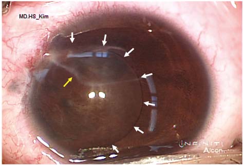
Fig. 1 Preoperative anterior segment photograph in an operative microscopic field of an inclusion cyst at POD 13 months after primary corneal closure. This photograph shows a linear corneal scar and a 6.0 mm fluid-filled epithelial inclusion cyst which occluded the pupil. White arrow=margin of epithelial inclusion cyst; Yellow arrow=previous corneal laceration scar.
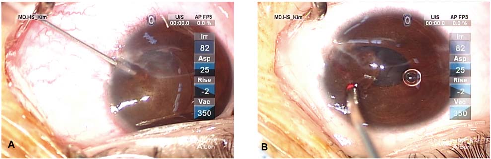
Fig. 2 Operative microscopic fields showing needle aspiration of a fluid-filled cyst (A) and Ab externo photocoagulation with an endolaser probe (B).
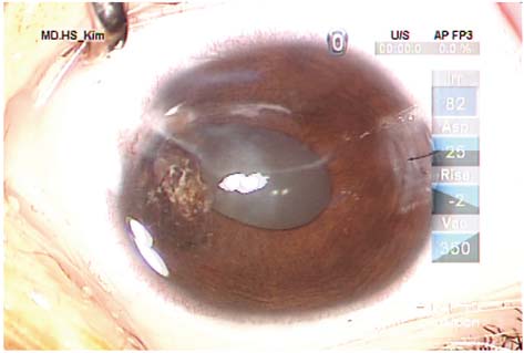
Fig. 3 Postoperative anterior segment photograph after needle aspiration and Ab externo laser photocoagulation treatment showing shrinkage of the large cyst.
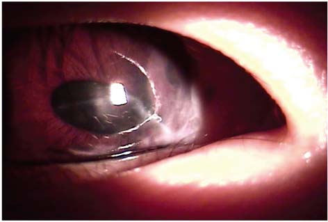
Fig. 4 Slit lamp photography showing disappearance of the large cyst in the right anterior chamber 6 months postoperatively. Best-corrected visual acuity was recovered to 20/40 after cyst aspiration and endolaser-mediated collapse of the cyst wall.
Reference
-
1. Perera C. Epithelium in the anterior chamber of the eye after operation and injury. Am J Ophthalmol. 1938. 21:605–611.2. Bennet T, D'amico RA. Epithelial inclusion cyst of iris after keratoplasty. Am J Ophthalmol. 1974. 77:87–89.3. Fiedman AH, Taterka HB, Henkind P. Epithelial implantation membrane on the iris surface following cataract extraction with a report of two cases. Am J Ophthalmol. 1971. 71:482–485.4. Verma L, Ray M, Sharma N, et al. Presumed epithelial inclusion cyst of the iris seven years after radial keratotomy. Cornea. 2002. 21:709–711.5. Haller JA, Stark WJ, Azab A, et al. Surgical approaches to the management of epithelial cysts. Trans Am Ophthalmol Soc. 2002. 100:79–84.6. Finger PT, McCormick SA, Lombardo J, et al. Epithelial inclusion cyst of the iris. Arch Ophthalmol. 1995. 113:777–780.7. Jester JV, Villasenor RA, Miyashiro J. Epithelial inclusion cyst following radial keratotomy. Arch Ophthalmol. 1983. 101:611–615.8. Tsai JC, Arrindel EL, O'Day DM. Needle aspiration and endodiathermy treatment of epithelial inclusion cyst of the iris. Am J Ophthalmol. 2001. 131:263–265.9. Shields JA. Primary cysts of the iris. Trans Am Ophthalmol Soc. 1981. 79:771–809.10. Shields JA, Klinc MW, Augsburger JJ. Primary iris cyst: a review of the literature and report of 62 cases. Br J Ophthalmol. 1984. 68:152–166.11. Lois N, Shields CL, Shields JA, Mercado G. Primary cysts of the iris pigment epithelium. Ophthalmology. 1998. 105:1879–1885.12. Shields JA, Shields CL, Lois N, Mercado G. Iris cysts in children: classification, incidence and management. Br J ophthalmol. 1999. 83:334–338.13. Alger EM. Large implantation cyst of the iris treated by aspiration and injection of iodine. Arch Ophthalmol. 1932. 7:984.14. Wilson W. Iris cyst treated with electrolysis. Br J Ophthalmol. 1964. 48:45–49.15. Maumenee AE, Paton D, Morse PH, Butner R. Review of 40 histologically proven cases of epithelial downgrowth following cataract extraction and suggested surgical management. Am J Ophthalmol. 1970. 69:598–603.16. Naumann GO, Rummelt V. Block excision of cystic and diffuse epithelial ingrowth of the anterior chamber: report on 32 consecutive patients. Arch Ophthalmol. 1992. 110:223–227.17. Cleasby GW. Photocoagulation of iris-ciliary-body epithelial cysts. Trans Am Acad Ophthalmol Otolaryngol. 1971. 75:638–642.18. Okun E, Mandell A. Photocoagulation as a treatment of epithelial cysts following cataract surgery. Trans Am Ophthalmol Soc. 1974. 72:170–183.19. Scholz RT, Kelley JS. Argon laser photocoagulation treatment of iris cysts following penetrating keratoplasty. Arch Ophthalmol. 1982. 100:926–927.20. Sugar J, Jampol LM, Goldberg MF. Argon laser destruction of anterior chamber implantation cysts. Ophthalmology. 1984. 91:1040–1044.21. Sihota R, Tiwari HK, Azad RV, Khosla PK. Photocoagulation of large iris cysts. Ann Ophthalmol. 1988. 20:470–472.
