Anterior Chamber Measurements by Pentacam and AS-OCT in Eyes With Normal Open Angles
- Affiliations
-
- 1Institute of Vision Research, Department of Ophthalmology, Yonsei University College of Medicine, Seoul, Korea. kcyeye@yuhs.ac
- 2Siloam Eye Hospital, Seoul, Korea.
- KMID: 1084211
- DOI: http://doi.org/10.3341/kjo.2008.22.4.242
Abstract
- PURPOSE: To assess the reproducibility and agreement of anterior chamber measurements between the Pentacam (PTC) and the Anterior segment optical coherence tomography (AOCT) in normal healthy eyes with open angle. METHODS: Prospective cross-sectional comparative case series. A total of 162 eyes of 81 healthy volunteers with normal open angle were included in this study. Anterior chamber angle (ACA) and anterior chamber depth (ACD) were measured with PTC and AOCT. Intra-observer variability and inter-methods agreement of both instruments for ACA and ACD were evaluated. RESULTS: Values of temporal and nasal ACA measured by two instruments were similar, and the results of ACD were also not significantly different between modalities (p>0.01). ACA and ACD measurements by PTC and AOCT showed good intra-observer and inter-method agreements (all >0.9). CONCLUSIONS: PTC and AOCT are presumed to be very useful for the anterior chamber angle examination. They may provide good images and quantitative data about the angle structures including ACA and ACD.
Keyword
MeSH Terms
-
Adolescent
Adult
Anterior Chamber/*anatomy & histology
Anterior Eye Segment
Cross-Sectional Studies
*Diagnostic Techniques, Ophthalmological
Female
Humans
Male
Observer Variation
Photography/*methods
Prospective Studies
Reproducibility of Results
Tomography, Optical Coherence/*methods
Trabecular Meshwork/anatomy & histology
Young Adult
Figure
-
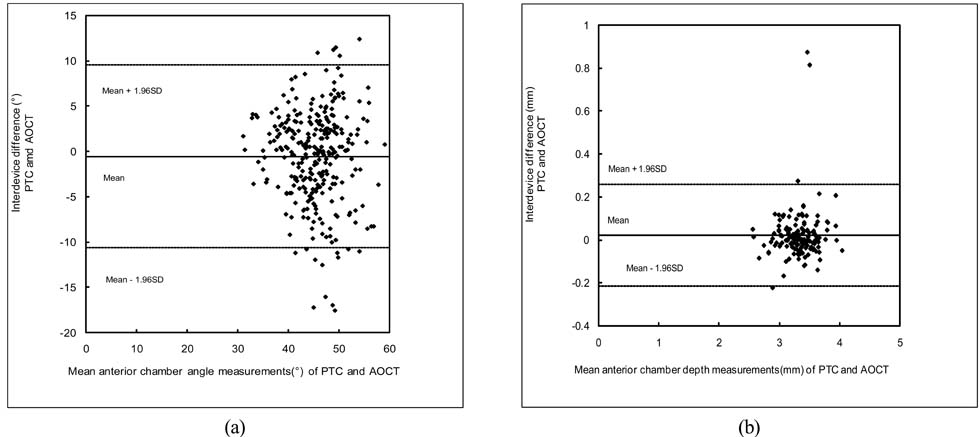
Fig. 1 Bland-Altman analysis for the intrer-methods agreement for Pentacam (PTC) and Anterior segment optical coherence tomography (AOCT). (a) Anterior chamber angle; (b) Anterior chamber depth.
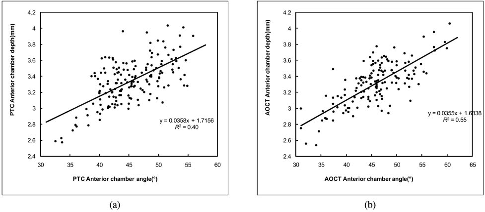
Fig. 2 Linear regression analysis between anterior chamber angle and anterior chamber depth for the Pentacam (PTC) (a) and the Anterior segment optical coherence tomography (AOCT) (b).
Cited by 4 articles
-
Comparison of Measurement of Anterior Segment Parameters Between Scheimpflug Camera and Ultrasound Biomicroscopy
Yong-sik Park, Hyunseok Ahn, Na Rae Kim, Kyoung Tak Ma, Samin Hong, Gong Je Seong, Chan Yun Kim
J Korean Ophthalmol Soc. 2009;50(12):1847-1852. doi: 10.3341/jkos.2009.50.12.1847.Comparison and Repeatability of Anterior Segment Parameters Obtained by Galilei and Slit-lamp Optical Coherence Tomography
Won Hyuk Lee, Young Hoon Hwang, Se Jong Kim, Sang Mok Lee, Chungkwon Yoo, Yong Yeon Kim, Joo Hwa Lee
J Korean Ophthalmol Soc. 2011;52(1):53-59. doi: 10.3341/jkos.2011.52.1.53.Comparison of Anterior Segment Measurements between Scheimpflug Camera and New Module of Optical Coherence Tomography
Jae Min Kim, Min Seok Kang, Kyung Hyun Jin
J Korean Ophthalmol Soc. 2018;59(7):613-621. doi: 10.3341/jkos.2018.59.7.613.Clinical Usefulness of UBM in the Sitting Position in Anterior Chamber Depth and Angle Measurements
Tae Gi Kim, Sung Woon Moon, Ji Ho Yang, Kyung Hyun Jin
J Korean Ophthalmol Soc. 2014;55(7):1007-1016. doi: 10.3341/jkos.2014.55.7.1007.
Reference
-
1. Bonomi L, Varotto A. Epidemiology of Angle-closure Glaucoma; Prevalence, Clinical Types, and Association with Peripheral Anterior Chamber Depth in the Egna-Neumarkt Galucoma Study. Opthalmology. 2000. 107:998–1003.2. Foster PJ, Johson GJ. Glaucoma in China-how big is the problem? Br J Ophthalmol. 2001. 85:1271–1272.3. Alsbrik PH. Anterior chamber depth and primary angle-closure glaucoma. I. An epidemiologic study in Greenland Eskimos. Acta Ophthalmol (Copenh). 1975. 53:89–104.4. Chew PT, Sung T. Primary angle-closure glaucoma in Asia. J Glaucoma. 2001. 10:Suppl. S7–S8.5. Dandona L, Dandona R, Mandal P, et al. Angle-closure glaucoma in an urban population in southern India. The Andhra Pradesh eye disease study. Ophthalmology. 2000. 107:1710–1716.6. Radhakrishnan S, Goldsmith J, Huang D, et al. Comparison of Optical Coherence Tomography and Ultrasound Biomicroscopy for Detection of Narrow Anterior Chamber Angles. Arch Ophthalmol. 2005. 123:1053–1059.7. Radhakrishnan S, Goldsmith J, Huang D, et al. Optical coherence tomography imaging of the anterior chamber angle. Ophthalmol Clin North Am. 2005. 18:375–381.8. Foster PJ, Devereux JG, Alsbirk PH, et al. Detection of gonioscopically occludable angles and primary angle closure glaucoma by estimation of limbal chamber depth in Asians: modified grading scheme. Br J Ophthalmol. 2000. 84:186–192.9. Foster PJ, Buhrmann R, Quigley HA, Johnson GJ. The definition and classification of glaucoma in prevalence surveys. Br J Ophthalmol. 2002. 86:238–242.10. Friedman DS. Who needs an iridotomy? Br J Ophthalmol. 2001. 85:1019–1021.11. Lackner B, Schimidger G, Skorpik C. Validity and Repeatability of Anterior Chamber Depth Measurements With Pentacam and Orbscan. Optom Vis Sci. 2005. 82:858–861.12. Bland JM, Altman DG. Statistical methods for assessing agreement between two methods of clinical measurement. Lancet. 1986. 1:307–310.13. Reddy AR, Pande MV, Finn P, El-Gogary H. Comparative estimation of anterior chamber depth by ultrasonography, Orbscan II, and IOLMaster. J Cataract Refract Surg. 2004. 30:1268–1271.14. Koranyi G, Lydahl E, Norrby S, Taube M. Anterior chamber depth measurement: a-scan versus optical methods. J Cataract Refract Surg. 2002. 28:243–247.15. Buehl W, Stojanac D, Sacu S, et al. Comparison of three methods of measuring corneal thickness and anterior chamber depth. Am J Ophthalmol. 2006. 141:7–12.
- Full Text Links
-
- Actions
-
Cited
- CITED
-
- Close
- Share
-
- Similar articles
-
- Detection of Occludable Angles with the Pentacam and the Anterior Segment Optical Coherence Tomography
- Clinical Usefulness of UBM in the Sitting Position in Anterior Chamber Depth and Angle Measurements
- Comparison of Measurement of Anterior Segment Parameters Between Scheimpflug Camera and Ultrasound Biomicroscopy
- Quantified Values of Anterior Chamber Depth and Angle Measurements Using Ultrasound Biomicroscopy and Topography
- Comparison of Corneal Measurement Values between Two Types of Topography
