Immersion Ultrasonography of Excised Nonpalpable Breast Lesion Specimens after Ultrasound-Guided Needle Localization
- Affiliations
-
- 1Department of Radiology, Korea University Ansan Hospital, Korea University College of Medicine, Gyeonggi-do, Korea. seoboky@korea.ac.kr
- 2Department of Radiology, Korea University Anam Hospital, Korea University College of Medicine, Seoul, Korea.
- 3Department of Radiology, Korea University Guro Hospital, Korea University College of Medicine, Seoul, Korea.
- 4Department of Pathology, Korea University Ansan Hospital, Korea University College of Medicine, Gyeonggi-do, Korea.
- 5Department of General Surgery, Korea University Ansan Hospital, Korea University College of Medicine, Gyeonggi-do, Korea.
- KMID: 1076456
- DOI: http://doi.org/10.3348/kjr.2008.9.4.312
Abstract
OBJECTIVE
Ultrasound-guided needle localization has been used prior to the surgical excision of nonpalpable breast lesions. The aim of the study was to assess the feasibility of the use of a saline immersion specimen ultrasound technique (immersion-US) to confirm the successful removal of breast lesions. MATERIALS AND METHODS: The devised immersion-US technique was used to examine the excised tissues of 72 ultrasound-guided needle localized breast lesions of 58 patients (34 benign lesions, 30 high-risk lesions and 8 malignant lesions). Freshly excised specimens were placed in a container filled with saline and one radiologist scanned the surgically excised specimens using a high-frequency linear transducer. We evaluated successful lesion removal and the qualities of the immersion-US images. Miss rates were determined by the use of postoperative ultrasound during follow-up. RESULTS: All 72 lesions were identified by the use of immersion-US and satisfactory or excellent quality images were obtained for most lesions (70/72, 97%). Five (7%) lesions were initially identified as incompletely excised, based on the immersion-US findings, and prompt re-excision was undertaken. Follow-up ultrasound examinations showed no residual mass in the surgical field in any patient. CONCLUSION: The immersion-US technique was found straightforward and efficient to perform. Immersion-US was able to determine whether nonpalpable breast lesions had been successfully excised after ultrasound-guided needle localization.
Keyword
MeSH Terms
Figure
-
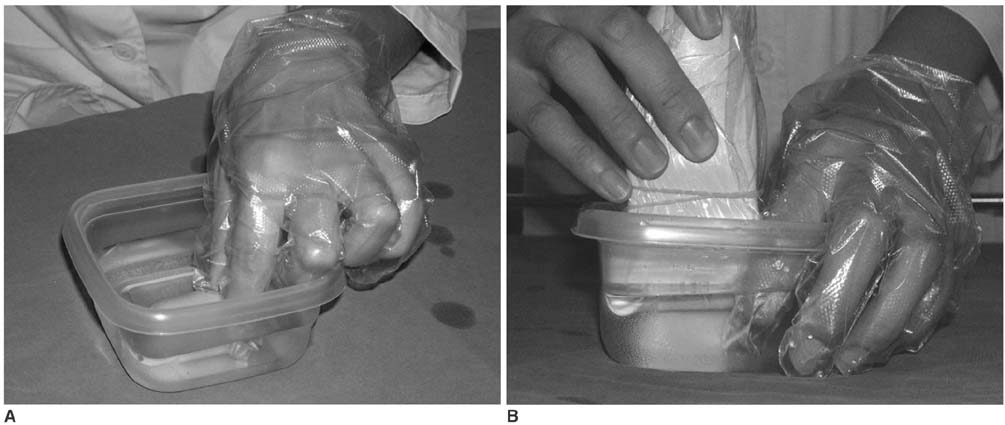
Fig. 1 Description of devised saline immersion technique for specimen US (immersion-US). Specimen is placed in plastic container filled with saline. With one hand holding specimen (A), sample is scanned with linear transducer with other hand (B).
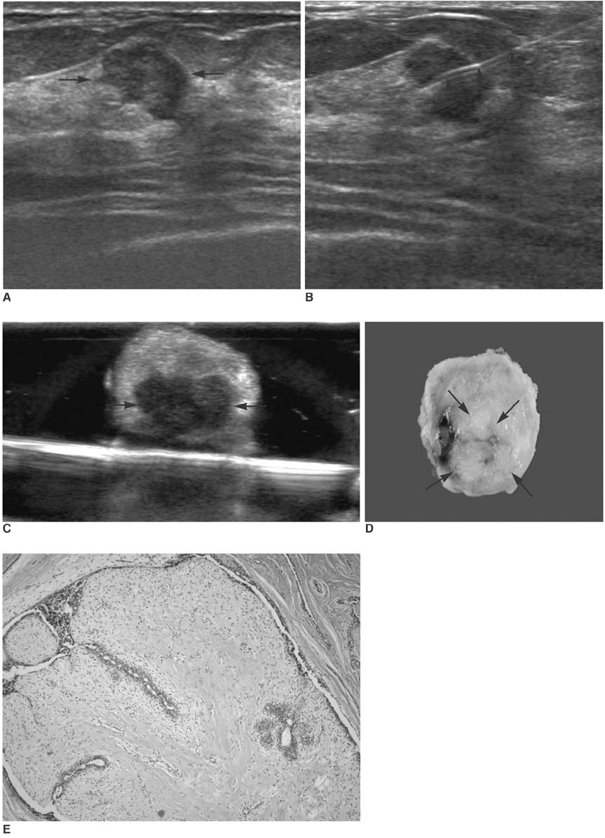
Fig. 2 36-year-old woman with fibroadenoma. A. US image shows 14 mm-sized, angular marginated, oval, hypoechoic mass (arrows). B. US-guided needle localization was performed for excision, and needle was directly passed through mass (arrowheads). C. After excision, immersion-US was performed and mass (arrows) was found to be successfully removed. D. Gross pathological examination shows whitish gray multilobulated solid mass (arrows). E. Microscopic pathological examination shows well-defined mass with fibrous stroma and elongated tubules (Hematoxylin & Eosin staining; original magnification, ×100).
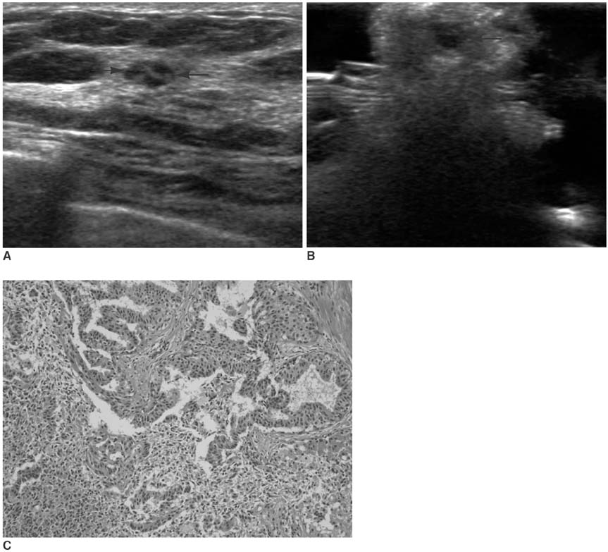
Fig. 3 44-year-old woman with intraductal papilloma. A. US image demonstrates 6-mm sized complex echoic mass (arrows). B. Immersion-US image shows complete mass excision (arrows). C. Microscopic pathologic examination demonstrates ductal epithelial papillary projections (Hematoxylin & Eosin staining; original magnification, ×200).
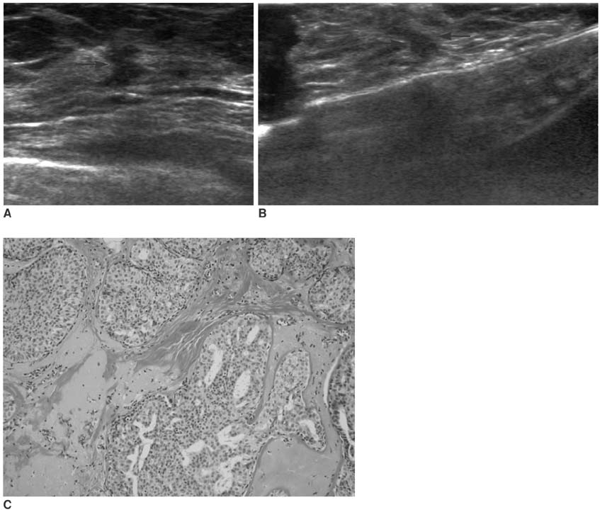
Fig. 4 46-year-old woman with ductal carcinoma in situ. A. US image shows 11-mm sized, spiculated irregularly shaped, isoechoic mass (arrows). B. Immersion-US image demonstrates successful mass excision (arrows). C. Microscopic pathological examination demonstrates cribriform and solid type, low-grade ductal carcinoma in situ (Hematoxylin & Eosin staining; original magnification, ×200).

Fig. 5 26-year-old woman with ductal carcinoma in situ and mucocele-like tumor. A. US shows 13-mm sized complex echoic mass (arrows). B. Immersion-US image demonstrates successful mass excision (arrows). C. Microscopic pathological examination demonstrates mucin pool with floating mucin producing malignant cells (arrows) (Hematoxylin & Eosin staining; original magnification, ×100).
Reference
-
1. Snider HC Jr, Morrison DG. Intraoperative ultrasound localization of nonpalpable breast lesions. Ann Surg Oncol. 1999. 6:308–314.2. American College of Radiology. BI-RADS Committee. . ACR BI-RADS® breast imaging and reporting data system: breast imaging atlas. 2003. Reston, VA: American College of Radiology.3. Ko K, Han BK, Jang KM, Choe YH, Shin JH, Yang JH, et al. The value of ultrasound-guided tattooing localization of nonpalpable breast lesions. Korean J Radiol. 2007. 8:295–301.4. Laing FC, Jeffrey RB, Minagi H. Ultrasound localization of occult breast lesions. Radiology. 1984. 151:795–796.5. Fornage BD, Ross MI, Singletary SE, Paulus DD. Localization of impalpable breast masses: value of sonography in the operating room and scanning of excised specimens. AJR Am J Roentgenol. 1994. 163:569–573.6. Mesurolle B, El-Khoury M, Hori D, Phancao JP, Kary S, Kao E, et al. Sonography of postexcision specimens of nonpalpable breast lesions: value, limitations, and description of a method. AJR Am J Roentgenol. 2006. 186:1014–1024.7. Harlow SP, Krag DN, Ames SE, Weaver DL. Intraoperative ultrasound localization to guide surgical excision of nonpalpable breast carcinoma. J Am Coll Surg. 1999. 189:241–246.8. Feld RI, Rosenberg AL, Nazarian LN, Needleman L, Lev-Toaff AS, Segal SR, et al. Intraoperative sonographic localization of breast masses. J Ultrasound Med. 2001. 20:959–966.9. Silverstein MJ, Lagios MD, Groshen S, Waisman JR, Lewinsky BS, Martino S, et al. The influence of margin width on local control of ductal carcinoma in situ of the breast. N Engl J Med. 1999. 340:1455–1461.
- Full Text Links
-
- Actions
-
Cited
- CITED
-
- Close
- Share
-
- Similar articles
-
- Needle Localization Biopsy of Nonpalpable Lesions of the Breast
- Usefulness of Ultrasound-Guided Automated Core Biopsy of Nonpalpable Breast Lesions
- US-Guided Preoperative Hook-Wire Localization of Nonpalpable Breast Lesions
- Feasibility of ultrasound-guided absorbable retaining thread needle localization for nonpalpable breast lesions
- The Results of Mammography and Ultrasound-Guided Localization Biopsy of Nonpalpable Breast Lesions, and the Differences between Them
