Cytotoxicity of gamma-ray in rat immature hippocampal neurons
- Affiliations
-
- 1Department of Veterinary Anatomy, College of Veterinary Medicine, Chonnam National University, Gwangju 500-757, Korea. moonc@chonnam.ac.kr
- 2Department of Veterinary Toxicology, College of Veterinary Medicine, Chonnam National University, Gwangju 500-757, Korea.
- 3Laboratory of Experimental Radiology, Research Center, Dongnam Institute of Radiological & Medical Science (DIRAMS), Busan 619-753, Korea.
- 4Department of Veterinary Anatomy, College of Veterinary Medicine and Applied Radiological Science Research Institute, Jeju National University, Jeju 690-756, Korea.
- KMID: 1067390
- DOI: http://doi.org/10.4142/jvs.2011.12.3.203
Abstract
- This in vitro study evaluated the detrimental effect of acute gamma (gamma)-irradiation on rat immature hippocampal neurons. Rat immature hippocampal neurons (0.5 day in vitro) were irradiated with 0~4 Gy gamma-rays. Cytotoxicity was analyzed using a lactate dehydrogenase release assay at 24 h after gamma-irradiation. Radiation-induced cytotoxicity in immature hippocampal neurons increased in a dose-dependent manner. Pre-treatments of pro-apoptotic caspase inhibitors and anti-oxidative substances significantly blocked gamma-irradiation-induced cytotoxicity in immature hippocampal neurons. The results suggest that the caspase-dependent cytotoxicity of gamma-rays in immature hippocampal cultured neurons may be caused by oxidative stress.
Keyword
MeSH Terms
-
Amifostine/pharmacology
Animals
Antioxidants/pharmacology
Caspase 3/metabolism/radiation effects
Catechin/analogs & derivatives/pharmacology
Cell Survival/radiation effects
Cells, Cultured/cytology/enzymology/*radiation effects
Dose-Response Relationship, Radiation
Female
*Gamma Rays
Hippocampus/cytology/enzymology/*radiation effects
L-Lactate Dehydrogenase/radiation effects
Neurons/cytology/enzymology/*radiation effects
Poly(ADP-ribose) Polymerases/drug effects
Pregnancy
Rats
Rats, Sprague-Dawley
Figure
-

Fig. 1 Double-immunofluorescent images of Ki-67 and nestin (A~D) and doublecortin (DCX) and glial fibrillary acidic protein (GFAP) (E~H) in 0.5 day in vitro-cultured hippocampal neurons. (A) Immunolabeling of Ki-67 (red). (B) Immuno-labeling of nestin (green). (C) 4',6-diamidino-2-phenylindole (DAPI) staining (blue). (D) Merged photograph. (E) Immunolabeling of DCX (red). (F) Immuno-labeling of GFAP (green). (G) DAPI staining (blue). (H) Merged photograph. Scale bars = 30 µm.
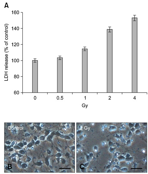
Fig. 2 Dose-dependent pattern of γ-ray-induced cytotoxicity in immature hippocampal neurons. (A) Lactate dehydrogenase (LDH) assay as an indicator of cytotoxicity of γ-rays (0~4 Gy) at 24 h after irradiation in hippocampal neurons of 0.5 DIV. (B and C) Differential interference contrast images revealed that cellular shrinkage and loss in irradiated cells (2 Gy) was increased compared with sham-irradiated controls. The data in (A) represent the mean ± SE, n = 3 cultures per condition. Scale bars = 30 µm.
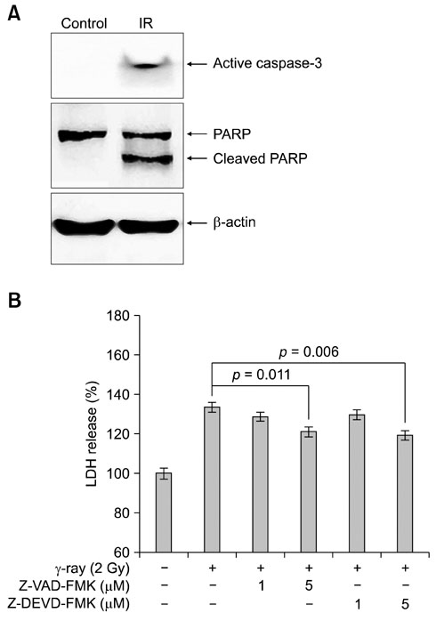
Fig. 3 Western blot analysis showing active caspase-3 and cleaved poly (ADP-ribose) polymerase (PARP) expression in immature hippocampal neurons with γ-irradiation (2 Gy). (A) Representative immunoblots for detection of active caspase-3 (17 kDa), PARP (118 kDa) and cleaved PARP (89 kDa) at 24 h after γ-irradiation (IR, 2 Gy). (B) 2 Gy γ-irradiated cells displayed significant increase of LDH in the medium. However, pre-treatment of the caspase-family inhibitor Z-VAD-FMK or the caspase-3 specific inhibitor Z-DEVD-FMK significantly inhibited release of LDH in the medium of 2 Gy-irradiated cells. The data represents the mean ± SE, n = 3 cultures per condition.
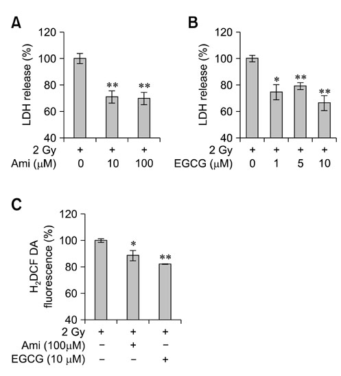
Fig. 4 The inhibitory effects of anti-oxidative substances on γ-ray-induced cytotoxicity and increase of intracellular reactive oxygen species (ROS) in immature hippocampal neurons. (A and B) Pre-treatments of anti-oxidative substances, amifostine (Ami) and epigallocatechin gallate (EGCG) at 30 min before irradiation significantly inhibited LDH release in immature hippocampal neurons with all of treated doses compared with that in 2 Gy-irradiated controls. (C) Pre-treatments of amifostine (100 µM) and EGCG (10 µM) significantly blocked cellular levels of ROS by 2 Gy-irradiation. The data represent the mean ± SE, n = 3 cultures per condition. *p < 0.05, **p < 0.01 vs. 2 Gy-irradiated controls.
Reference
-
1. Bayer SA. Cellular aspects of brain development. Neurotoxicology. 1989. 10:307–320.2. Fike JR, Rosi S, Limoli CL. Neural precursor cells and central nervous system radiation sensitivity. Semin Radiat Oncol. 2009. 19:122–132.
Article3. Fukuda H, Fukuda A, Zhu C, Korhonen L, Swanpalmer J, Hertzman S, Leist M, Lannering B, Lindholm D, Björk-Eriksson T, Marky I, Blomgren K. Irradiation-induced progenitor cell death in the developing brain is resistant to erythropoietin treatment and caspase inhibition. Cell Death Differ. 2004. 11:1166–1178.
Article4. Gobbel GT, Bellinzona M, Vogt AR, Gupta N, Fike JR, Chan PH. Response of postmitotic neurons to X-irradiation: implications for the role of DNA damage in neuronal apoptosis. J Neurosci. 1998. 18:147–155.
Article5. Hall P, Adami HO, Trichopoulos D, Pedersen NL, Lagiou P, Ekbom A, Ingvar M, Lundell M, Granath F. Effect of low doses of ionising radiation in infancy on cognitive function in adulthood: Swedish population based cohort study. BMJ. 2004. 328:19.
Article6. Hays SR, Li X, Kimler BF. Is there an adaptive response to radiation in the developing brain of the fetal rat? Radiat Res. 1993. 136:293–296.
Article7. Kim JS, Lee HJ, Kim JC, Kang SS, Bae CS, Shin T, Jin JK, Kim SH, Wang H, Moon C. Transient impairment of hippocampus-dependent learning and memory in relatively low-dose of acute radiation syndrome is associated with inhibition of hippocampal neurogenesis. J Radiat Res (Tokyo). 2008. 49:517–526.
Article8. McDonough JH, Mele PC, Franz CG. Comparison of behavioral and radioprotective effects of WR-2721 and WR-3689. Pharmacol Biochem Behav. 1992. 42:233–243.
Article9. Moore AH, Olschowka JA, Williams JP, Paige SL, O'Banion MK. Radiation-induced edema is dependent on cyclooxygenase 2 activity in mouse brain. Radiat Res. 2004. 161:153–160.
Article10. Packer RJ, Sutton LN, Atkins TE, Radcliffe J, Bunin GR, D'Angio G, Siegel KR, Schut L. A prospective study of cognitive function in children receiving whole-brain radiotherapy and chemotherapy: 2-year results. J Neurosurg. 1989. 70:707–713.
Article11. Shirai K, Mizui T, Suzuki Y, Kobayashi Y, Nakano T, Shirao T. Differential effects of x-irradiation on immature and mature hippocampal neurons in vitro. Neurosci Lett. 2006. 399:57–60.
Article12. Song MS, Kim JS, Yang M, Kim SH, Kim JC, Park SH, Shin T, Moon C. Gamma-ray susceptibility of immature and mature hippocampal cultured cells. J Vet Med Sci. 2010. 72:605–609.
Article13. Verderio C, Coco S, Pravettoni E, Bacci A, Matteoli M. Synaptogenesis in hippocampal cultures. Cell Mol Life Sci. 1999. 55:1448–1462.
Article
- Full Text Links
-
- Actions
-
Cited
- CITED
-
- Close
- Share
-
- Similar articles
-
- Comparison of the GABAergic currents associated with midazolam and propofol in rat hippocampal neurons
- C-fos mRNA Expression in Rat Hippocampal Neurons by Antidepressant Drugs
- Effect of Developmental Lead Exposure on the Expression of Hippocampal NMDA Receptor Subunit mRNA
- Effect about Neurite Extension of FS390, an Inhibitor of Exocytosis in Rat Hippocampal Neurons and PC12 Cells
- Recurrent Early-Life Seizures and Changes in GABAA Receptors Expression in Hippocampus
