J Vet Sci.
2006 Jun;7(2):199-201. 10.4142/jvs.2006.7.2.199.
Mediastinal lymphoma in a young Turkish Angora cat
- Affiliations
-
- 1Department of Veterinary Internal Medicine, College of Veterinary Medicine, Seoul National University, Seoul 151-742, Korea. cyhwang@snu.ac.kr
- 2Department of Veterinary Clinical Pathology, College of Veterinary Medicine, Seoul National University, Seoul 151-742, Korea.
- 3Department of Veterinary Pathology, College of Veterinary Medicine, Seoul National University, Seoul 151-742, Korea.
- KMID: 1059209
- DOI: http://doi.org/10.4142/jvs.2006.7.2.199
Abstract
- An 8-month old intact male Turkish Angora cat was referred to the Veterinary Medical Teaching Hospital (VMTH), Seoul National University, for an evaluation of anorexia and severe dyspnea. The thoracic radiographs revealed significant pleural effusion. A cytology evaluation of the pleural fluid strongly suggested a lymphoma containing variable sized lymphocytes with frequent mitotic figures and prominent nucleoli. The feline leukemia virus and feline immunodeficiency virus tests were negative. The cat was euthanized at his owner's request and a necropsy was performed. A mass was detected on the mediastinum and lung lobes. A histopathology evaluation confirmed the mass to be a lymphoma. Immunohistochemistry revealed the mass to be CD3 positive. In conclusion, the cat was diagnosed as a T-cell mediastinal lymphoma.
MeSH Terms
Figure
-

Fig. 1 Cytologic smear of pleural effusion of the cat. Note the neoplastic large lymphocytes with the diameter of nuclei more than 3 times that of RBC and a large central nucleolus. There is one mitotic figure in the center. Wright stain, ×100.
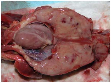
Fig. 2 Gross lesion of the large mass from thorax necropsy of a cat diagnosed mediastinal lymphoma. Note the abnormal large mass around the heart instead of normal lung structures.

Fig. 3 Histological section of feline mediastinal mass, diagnosed as a mediastinal lymphoma. Note compact sheet of monomorphic lymphoid cells. H&E stain, ×150.
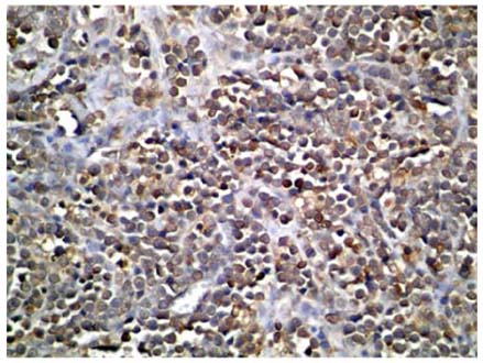
Fig. 4 Immunohistochemical staining for CD3 detection of feline mediastinal lymphoma. ×40.
Reference
-
1. Antony SM, Gregory KO. Gregory KO, Antony SM, editors. Lymphoma. Feline Oncology. 2001. Trenton, Jackson: Veterinary Learning Systems;191–219.2. Court EA, Watson AD, Peaston AE. Retrospective study of 60 cases of feline lymphosarcoma. Aust Vet J. 1997. 75:424–427.
Article3. Crighton GW. Clinical aspects of lymphosarcoma in the cat. Vet Rec. 1968. 83:122–126.4. Gabor LJ, Canfield PJ, Malik R. Immunophenotypical and histological characterization of 109 cases of feline lymphosarcoma. Aust Vet J. 1999. 77:436–441.
Article5. Gabor LJ, Marlik R, Canfield PJ. Clinical and anatomical features of lymphosarcoma in 118 cats. Aust Vet J. 1998. 76:725–732.
Article6. Gruffydd-Jones TJ, Gaskell CJ, Gibbs C. Clinical and radiological features of anterior mediastinal lymphosarcoma in the cat: a review of 30 cases. Vet Rec. 1979. 104:304–307.
Article7. Louwerens M, London CA, Pederson NC, Lyons LA. Feline lymphoma in the post-feline leukemia virus era. J Vet Intern Med. 2005. 19:329–335.
Article8. Peaston AE, Maddison JE. Efficacy of doxorubicin as an induction agent for cats with lymphosarcoma. Aust Vet J. 1999. 77:442–444.
Article9. Takahashi R, Goto N, Ishii H, Ogiso Y, Saegusa J. Pathological observations of natural cases of feline lymphosarcomatosis. Nihon Juigaku Zasshi. 1974. 36:163–173.
Article10. Vail DM, Moore AS, Ogilvie GK, Volk LM. Feline lymphoma (145 cases): proliferation indices, cluster of differentiation 3 immunoreactivity, and their association with prognosis in 90 cats. J Vet Intern Med. 1998. 12:349–354.
Article
