Atypical Developmental Venous Anomaly Associated with Single Arteriovenous Fistula and Intracerebral Hemorrhage: a Case Demonstrated by Superselective Angiography
- Affiliations
-
- 1Department of Radiology, Chungbuk National University Hospital, Chungbuk 361-711, Korea. shcha@chungbuk.ac.kr
- 2Department of Neurosurgery, Chungbuk National University Hospital, Chungbuk 361-711, Korea.
- KMID: 1058802
- DOI: http://doi.org/10.3348/kjr.2012.13.1.107
Abstract
- We present a case of developmental venous anomaly associated with arteriovenous fistula supplied by a single arterial feeder adjacent to a large acute intracerebral hemorrhage. The arteriovenous fistula was successfully obliterated by superselective embolization while completely preserving the developmental venous anomaly. Two similar cases, including superselective angiographic findings, have been reported in the literature; however, we describe herein superselective angiographic findings in more detail and demonstrate the arteriovenous shunt more clearly than the previous reports. In addition, a literature review was performed to discuss the association of a developmental venous anomaly with vascular lesions.
Keyword
MeSH Terms
Figure
-
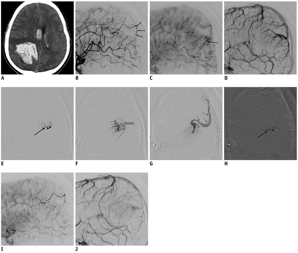
Fig. 1 18-year-old male with atypical developmental venous anomaly. A. Initial precontrast brain CT image showing large intracerebral hemorrhage in right parietal lobe and intraventricular hemorrhage in bilateral lateral ventricles. B. Right internal carotid angiogram of lateral image showing arterial pedicle (arrows) from pericallosal branch of right anterior cerebral artery. C. Late arterial phase angiogram showing early venous drainage (arrow). D. Developmental venous anomaly visualized in right parietal lobe in venous phase. E-G. Serial microangiogram clearly demonstrating arterial pedicle (arrowheads), site of arteriovenous fistula (large arrow) and early venous drainage (small arrows). Venous drainage from arteriovenous fistula shares venous channel of developmental venous anomaly showed in D and tip of microcatheter (open arrow) is also seen. H. Cast of NBCA-Lipiodol is located in distal arterial pedicle (arrowhead), arteriovenous fistula (large arrow), and venous channel just distal to arteriovenous fistula (small arrow). I, J. Late arterial phase lateral projection image (I) showing delayed flow in pedicle along with no arteriovenous shunt in post-embolization angiogram. Developmental venous anomaly still persists in venous phase (J) after embolization.
Cited by 2 articles
-
Relationship between Abnormal Hyperintensity on T2-Weighted Images Around Developmental Venous Anomalies and Magnetic Susceptibility of Their Collecting Veins:
In-Vivo Quantitative Susceptibility Mapping Study
Yangsean Choi, Jinhee Jang, Yoonho Nam, Na-Young Shin, Hyun Seok Choi, So-Lyung Jung, Kook-Jin Ahn, Bum-soo Kim
Korean J Radiol. 2019;20(4):662-670. doi: 10.3348/kjr.2018.0685.Multimodal Imaging Follow-up of a Thrombosed Developmental Venous Anomaly: CT, CT Angiography and Digital Subtraction Angiography
Kyung Sik Yi, Sang-Hoon Cha, Kyung Soo Min
Neurointervention. 2013;8(2):120-124. doi: 10.5469/neuroint.2013.8.2.120.
Reference
-
1. Abe T, Singer RJ, Marks MP, Norbash AM, Crowley RS, Steinberg GK. Coexistence of occult vascular malformations and developmental venous anomalies in the central nervous system: MR evaluation. AJNR Am J Neuroradiol. 1998. 19:51–57.2. Hussain JZ, Ray A, Hughes DG, Leggate JR. Complex developmental venous anomaly of the brain. Acta Neurochir (Wien). 2002. 144:501–504.3. Mullan S, Mojtahedi S, Johnson DL, Macdonald RL. Cerebral venous malformation-arteriovenous malformation transition forms. J Neurosurg. 1996. 85:9–13.4. Sirin S, Kahraman S, Gocmen S, Erdogan E. A rare combination of a developmental venous anomaly with a varix. Case report. J Neurosurg Pediatr. 2008. 1:156–159.5. Aksoy FG, Gomori JM, Tuchner Z. Association of intracerebral venous angioma and true arteriovenous malformation: a rare, distinct entity. Neuroradiology. 2000. 42:455–457.6. Lindquist C, Guo WY, Karlsson B, Steiner L. Radiosurgery for venous angiomas. J Neurosurg. 1993. 78:531–536.7. Fierstien SB, Pribram HW, Hieshima G. Angiography and computed tomography in the evaluation of cerebral venous malformations. Neuroradiology. 1979. 17:137–148.8. Awad IA, Robinson JR Jr, Mohanty S, Estes ML. Mixed vascular malformations of the brain: clinical and pathogenetic considerations. Neurosurgery. 1993. 33:179–188. discussion 188.9. Meyer B, Stangl AP, Schramm J. Association of venous and true arteriovenous malformation: a rare entity among mixed vascular malformations of the brain. Case report. J Neurosurg. 1995. 83:141–144.10. Hirata Y, Matsukado Y, Nagahiro S, Kuratsu J. Intracerebral venous angioma with arterial blood supply: a mixed angioma. Surg Neurol. 1986. 25:227–232.11. Fok KF, Holmin S, Alvarez H, Ozanne A, Krings T, Lasjaunias PL. Spontaneous intracerebral hemorrhage caused by an unusual association of developmental venous anomaly and arteriovenous malformation. Interv Neuroradiol. 2006. 12:113–121.12. Oran I, Kiroglu Y, Yurt A, Ozer FD, Acar F, Dalbasti T, et al. Developmental venous anomaly (DVA) with arterial component: a rare cause of intracranial haemorrhage. Neuroradiology. 2009. 51:25–32.13. Im SH, Han MH, Kwon BJ, Ahn JY, Jung C, Park SH, et al. Venous-predominant parenchymal arteriovenous malformation: a rare subtype with a venous drainage pattern mimicking developmental venous anomaly. J Neurosurg. 2008. 108:1142–1147.14. Komiyama M, Yamanaka K, Iwai Y, Yasui T. Venous angiomas with arteriovenous shunts: report of three cases and review of the literature. Neurosurgery. 1999. 44:1328–1334. discussion 1334-1325.15. Kurita H, Sasaki T, Tago M, Kaneko Y, Kirino T. Successful radiosurgical treatment of arteriovenous malformation accompanied by venous malformation. AJNR Am J Neuroradiol. 1999. 20:482–485.
- Full Text Links
-
- Actions
-
Cited
- CITED
-
- Close
- Share
-
- Similar articles
-
- Repeated Intracerebral Hemorrhage from Developmental Venous Anomaly Alone
- Traumatic Carotid-cavernous Fistula Bringing about Intracerebral Hemorrhage
- Transcatheter arterial embolization for congenital renal arteriovenous fistula
- Developmental Venous Anomaly Presenting Intracranial Hemorrhage without Associated Vascular Anomaly
- Multimodal Imaging Follow-up of a Thrombosed Developmental Venous Anomaly: CT, CT Angiography and Digital Subtraction Angiography
