Laser Peripheral Iridotomy with Iridoplasty in Primary Angle Closure Suspect: Anterior Chamber Analysis by Pentacam
- Affiliations
-
- 1HanGil Eye Hospital, Incheon, Korea. deskshot@naver.com
- KMID: 1018413
- DOI: http://doi.org/10.3341/kjo.2011.25.4.252
Abstract
- PURPOSE
To compare conventional laser peripheral iridotomy (LPI) and LPI combined with laser peripheral iridoplasty in eyes with primary angle closure suspect (PACS) by assessment of anterior chamber dimensional changes using a Pentacam.
METHODS
Forty-eight eyes of 24 subjects with bilateral PACS were recruited consecutively. Each eye was randomly allocated to treatment with conventional LPI, argon LPI only, or LPI plus iridoplasty, which consisted of simultaneous argon LPI and peripheral iridoplasty. Anterior chamber measurements were performed on each eye using a Pentacam, both before and after treatment. Mean anterior chamber depth (ACD), anterior chamber volume (ACV), and anterior chamber angle were measured, and topographic ACD analysis was performed. Results were compared between the two treatment groups.
RESULTS
After treatment with either conventional LPI or LPI plus iridoplasty, the mean ACD and ACV increased significantly. Topographic ACD analysis revealed that the mid-to-peripheral ACD increase was significantly greater in the LPI plus iridoplasty group than in eyes treated with conventional LPI. Intraocular pressure changes and post-LPI complications did not differ between the groups.
CONCLUSIONS
Compared with conventional LPI, our study showed that LPI plus iridoplasty improved the mid-to-peripheral ACD increase. This procedure may have a role as an adjunct for reducing angle closure by simultaneously eliminating pupillary and non-pupillary block components.
Keyword
MeSH Terms
-
Adult
Aged
Anterior Chamber/*pathology/surgery
Diagnostic Techniques, Ophthalmological/*instrumentation
Equipment Design
Female
Follow-Up Studies
Glaucoma, Angle-Closure/pathology/physiopathology/*surgery
Gonioscopy
Humans
Intraocular Pressure
Iridectomy/*methods
Iris/pathology/*surgery
Laser Therapy/*methods
Lasers, Solid-State
Male
Middle Aged
Prospective Studies
Tonometry, Ocular
Figure
-

Fig. 1 Conventional laser peripheral iridotomy (LPI) and the LPI combined with iridoplasty technique. (A) Conventional LPI refers to LPI alone. (B) The LPI combined with iridoplasty approach consists of LPI followed by laser peripheral iridoplasty administered in the same session. Yellow circle: LPI site, cyanine-blue circle: laser peripheral iridoplasty site; about 20 burn spots are placed alongside the limbus.
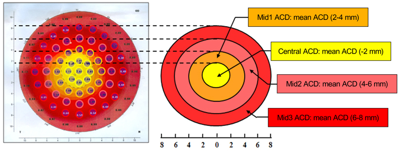
Fig. 2 Advanced anterior chamber depth (ACD) topographic parameters derived from routine ACD data from the Pentacam output. Central ACD is the average of ACD values at nine topographic points within 2 mm from the center of the eye; Mid1 ACD is the average of ACD measurements at 12 topographic points within 2 to 4 mm from the eye center; Mid2 ACD is the average of ACD values at 16 topographic points within 4 to 6 mm from the center of the eye; Mid3 ACD is the average of ACD measurements at 20 topographic points within 6 to 8 mm from the eye center.
Reference
-
1. European Glaucoma Society. Terminology and guidelines for glaucoma. 2008. 3rd ed. Savona: Dogma.2. Shen SY, Wong TY, Foster PJ, et al. The prevalence and types of glaucoma in malay people: the Singapore Malay Eye Study. Invest Ophthalmol Vis Sci. 2008. 49:3846–3851.3. Casson RJ, Newland HS, Muecke J, et al. Prevalence of glaucoma in rural Myanmar: the Meiktila Eye Study. Br J Ophthalmol. 2007. 91:710–714.4. Thomas R, George R, Parikh R, et al. Five year risk of progression of primary angle closure suspects to primary angle closure: a population based study. Br J Ophthalmol. 2003. 87:450–454.5. Thomas R, Parikh R, Muliyil J, Kumar RS. Five-year risk of progression of primary angle closure to primary angle closure glaucoma: a population-based study. Acta Ophthalmol Scand. 2003. 81:480–485.6. Ang MH, Baskaran M, Kumar RS, et al. National survey of ophthalmologists in Singapore for the assessment and management of asymptomatic angle closure. J Glaucoma. 2008. 17:1–4.7. Pandav SS, Kaushik S, Jain R, et al. Laser peripheral iridotomy across the spectrum of primary angle closure. Can J Ophthalmol. 2007. 42:233–237.8. He M, Friedman DS, Ge J, et al. Laser peripheral iridotomy in primary angle-closure suspects: biometric and gonioscopic outcomes: the Liwan Eye Study. Ophthalmology. 2007. 114:494–500.9. Kumar RS, Baskaran M, Chew PT, et al. Prevalence of plateau iris in primary angle closure suspects an ultrasound biomicroscopy study. Ophthalmology. 2008. 115:430–434.10. Kumar RS, Tantisevi V, Wong MH, et al. Plateau iris in Asian subjects with primary angle closure glaucoma. Arch Ophthalmol. 2009. 127:1269–1272.11. Pavlin CJ, Ritch R, Foster FS. Ultrasound biomicroscopy in plateau iris syndrome. Am J Ophthalmol. 1992. 113:390–395.12. Ritch R. Plateau iris is caused by abnormally positioned ciliary processes. J Glaucoma. 1992. 1:23–26.13. Ritch R, Tham CC, Lam DS. Argon laser peripheral iridoplasty (ALPI): an update. Surv Ophthalmol. 2007. 52:279–288.14. Ritch R, Tham CC, Lam DS. Long-term success of argon laser peripheral iridoplasty in the management of plateau iris syndrome. Ophthalmology. 2004. 111:104–108.15. Sassani JW, Ritch R, McCormick S, et al. Histopathology of argon laser peripheral iridoplasty. Ophthalmic Surg. 1993. 24:740–745.16. Van Herick W, Shaffer RN, Schwartz A. Estimation of width of angle of anterior chamber. Incidence and significance of the narrow angle. Am J Ophthalmol. 1969. 68:626–629.17. Boker T, Sheqem J, Rauwolf M, Wegener A. Anterior chamber angle biometry: a comparison of Scheimpflug photography and ultrasound biomicroscopy. Ophthalmic Res. 1995. 27:Suppl 1. 104–109.18. Pavlin CJ, Harasiewicz K, Foster FS. Ultrasound biomicroscopy of anterior segment structures in normal and glaucomatous eyes. Am J Ophthalmol. 1992. 113:381–389.19. Kurita N, Mayama C, Tomidokoro A, et al. Potential of the pentacam in screening for primary angle closure and primary angle closure suspect. J Glaucoma. 2009. 18:506–512.20. Rabsilber TM, Khoramnia R, Auffarth GU. Anterior chamber measurements using Pentacam rotating Scheimpflug camera. J Cataract Refract Surg. 2006. 32:456–459.
- Full Text Links
-
- Actions
-
Cited
- CITED
-
- Close
- Share
-
- Similar articles
-
- Effectiveness of Argon Laser Peripheral Iridoplasty in the Treatment of Severe Acute Angle-Closure Glaucoma
- Effect of Combined Argon Laser Peripheral Iridoplasty and Laser Iridotomy in Primary Angle-Closure Glaucoma
- Long-term Outcome of Anterior Chamber Parameters after Laser Iridotomy and Iridoplasty in Primary Angle Closure Glaucoma
- Effect of YAG Laser Iridotomy on IOP in Chronic Angle-closure Glaucoma
- Change in Angle Parameters Measured by Anterior Segment Optical Coherence Tomography after Laser Peripheral Iridotomy Alone versus Laser Peripheral Iridotomy and Argon Laser Peripheral Iridoplasty
