Korean J Ophthalmol.
2011 Jun;25(3):210-213. 10.3341/kjo.2011.25.3.210.
Neurotrophic Corneal Ulcer Development Following Cataract Surgery with a Limbal Relaxing Incision
- Affiliations
-
- 1Department of Ophthalmology, East-West Neo Medical Center, Kyung Hee University School of Medicine, Seoul, Korea.
- 2Department of Ophthalmology, Seoul Paik Hospital, Inje University College of Medicine, Seoul, Korea.
- 3Department of Ophthalmology and Visual Science, College of Medicine, The Catholic University of Korea, Seoul, Korea. chungsh@catholic.ac.kr
- KMID: 1010031
- DOI: http://doi.org/10.3341/kjo.2011.25.3.210
Abstract
- A 60-year-old man with bilateral corneal opacity underwent cataract extraction surgery involving the use of a limbal relaxing incision in his left eye. He had lower lid ectropion and lagophthalmos in both eyes. Eleven days after the surgery, a slit-lamp examination revealed a neurotrophic corneal ulcer with a punch-out epithelial defect and rolled edges at the center of the pre-existing corneal opacity. The patient was treated with sodium hyaluronate, autologous serum, and oral doxycycline. Six weeks after the surgery an improvement in corneal sensation was observed and the neurotrophic corneal ulcer subsequently healed over the course of one year. In this report, we present a case of neurotrophic keratitis that occurred after performing cataract surgery concurrent with a limbal relaxing incision. As such, we suggest that limbal relaxing incisions should be performed cautiously in patients with causative risk factors for corneal hypesthesia.
MeSH Terms
Figure
-
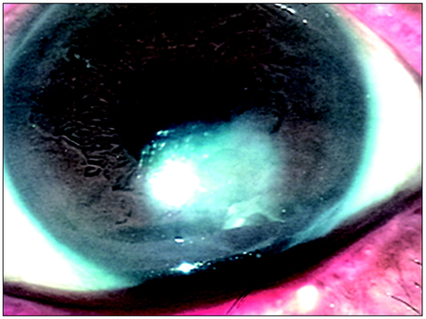
Fig. 1 Slit-lamp biomicroscopy on postoperative day 11. Note the punch-out epithelial defect (1.5 × 1.5 mm) with a border of hazy epithelium and stromal edema on the pre-existing corneal opacity, consistent with clinical stage 2 neurotrophic keratitis.
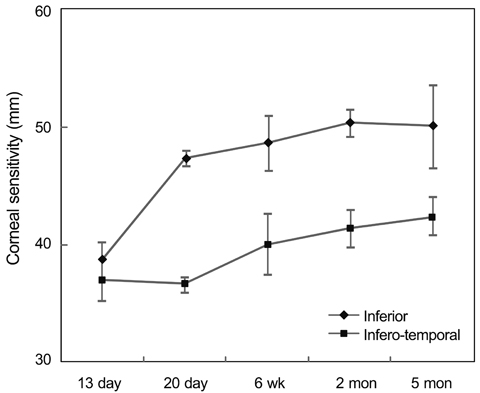
Fig. 2 The postoperative change in the mean corneal sensitivity. These data show the sensitivity of the inferior and infero-temporal cornea over the span of the first five months following surgery.
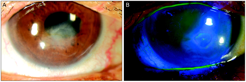
Fig. 3 Slit-lamp biomicroscopy at 10 months post-surgery. Note the oval-shaped epithelial thinning (1.5 × 1.5 mm) (A) and some erosion of the pre-existing corneal opacity (B).
Reference
-
1. Mamalis N. Correction of astigmatism during cataract surgery. J Cataract Refract Surg. 2009. 35:403–404.2. Bayramlar HH, Dağlioğlu MC, Borazan M. Limbal relaxing incisions for primary mixed astigmatism and mixed astigmatism after cataract surgery. J Cataract Refract Surg. 2003. 29:723–728.3. Kim HJ, Kim JW, Ahn JE. Limbal relaxing incision combined with cataract surgery reducing preoperative corneal astigmatism. J Korean Ophthalmol Soc. 2002. 43:1892–1900.4. Davis EA, Dohlman CH. Neurotrophic keratitis. Int Ophthalmol Clin. 2001. 41:1–11.5. John T. Corneal sensation after small incision, sutureless, one-handed phacoemulsification. J Cataract Refract Surg. 1995. 21:425–428.6. Kohlhaas M, Stahlhut O, Tholuck J, Richard G. Development of corneal sensitivity after phacoemulsification with scleral tunnel incision. Klin Monbl Augenheilkd. 1997. 211:32–36.7. Bonini S, Rama P, Olzi D, Lambiase A. Neurotrophic keratitis. Eye (Lond). 2003. 17:989–995.8. Jeng BH, Dupps WJ Jr. Autologous serum 50% eyedrops in the treatment of persistent corneal epithelial defects. Cornea. 2009. 28:1104–1108.9. Kohlhaas M. Corneal sensation after cataract and refractive surgery. J Cataract Refract Surg. 1998. 24:1399–1409.10. Lyne A. Corneal sensitivity after surgery. Trans Ophthalmol Soc U K. 1982. 102(pt 2):302–305.11. Khanal S, Tomlinson A, Esakowitz L, et al. Changes in corneal sensitivity and tear physiology after phacoemulsification. Ophthalmic Physiol Opt. 2008. 28:127–134.12. Kim YM, Kim SW, Kim TI, et al. The change of corneal sensitivity and recovery of corneal nerve after cataract surgery. J Korean Ophthalmol Soc. 2007. 48:13–18.13. Quinto GG, Campos M, Behrens A. Autologous serum for ocular surface diseases. Arq Bras Oftalmol. 2008. 71:6 Suppl. 47–54.14. Matsumoto Y, Dogru M, Goto E, et al. Autologous serum application in the treatment of neurotrophic keratopathy. Ophthalmology. 2004. 111:1115–1120.
- Full Text Links
-
- Actions
-
Cited
- CITED
-
- Close
- Share
-
- Similar articles
-
- Changes in Corneal Sensitivity after Cataract Surgery
- Limbal Relaxing Incision Combined with Cataract Surgery Reducing Preoperative Corneal Astigmatism
- The Short-Term Effect of Limbal Relaxing Incision and Compression Suture on Post-Penetrating Keratoplasty Astigmatism
- Mid Limbal Incision vs Scleral Pocket Incision in Cataract Surgery
- The Short Term Effects of a Single Limbal Relaxing Incision Combined with Clear Corneal Incision
