Korean J Ophthalmol.
2011 Jun;25(3):174-177. 10.3341/kjo.2011.25.3.174.
Quantitative Analysis of Optic Disc Color
- Affiliations
-
- 1Department of Ophthalmology, Kim's Eye Hospital, Konyang University College of Medicine, Seoul, Korea. yhsohn@kimeye.com
- KMID: 1010024
- DOI: http://doi.org/10.3341/kjo.2011.25.3.174
Abstract
- PURPOSE
To evaluate the reproducibility of ImageJ software in analyzing the color of the optic disc.
METHODS
One hundred twelve normal participants (56 males and 56 females) were enrolled in this study. The image of the optic disc was taken using Kowa digital disc photo-graphy, and the gray scales of the nasal rim (NR), brightest cupping center (BCC) and largest inferior retinal vein (IRV) were calculated using histogram in ImageJ. Three different observers calculated the gray scales three separate times. Reproducibility was assessed using the interclass correlation coefficient (ICC).
RESULTS
The mean age of the participants was 50.6 years old (range, 11 to 82 years). The mean gray scales of the nasal rim were 91.81, 94.91, and 93.24; those of the brightest cupping center were 174.84, 179.94, and 177.76; and those of the largest inferior retinal vein were 61.85, 53.48, and 56.73 for observers 1, 2, and 3, respectively. Inter-observer reproducibility for NR, BCC and IRV was considered good based upon ICC values of 0.944, 0.860, and 0.789 for observers 1, 2, and 3, respectively. Significant age-related differences between the values of the brightest cupping center were noted, and the gray scale score was decreased in the older participants (p < 0.001).
CONCLUSIONS
The gray scale of the brightest cupping center diminished with age. ImageJ can be a useful objective tool with high reproducibility in the analysis of optic disc color.
Keyword
MeSH Terms
Figure
-
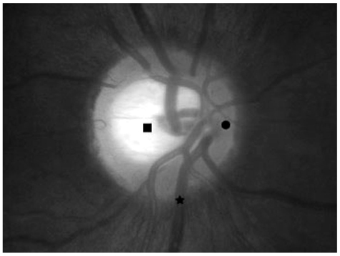
Fig. 1 Estimated points are shown in the photographs of the optic disc head. ● The nasal rim at the point of nine o'clock from the disc center. ■ The brightest cupping center is nearly the center of cupping where the color is brightest. ★ The largest inferior retinal vein is the point of the inferior retinal vein that comes in contact with the disc margin.
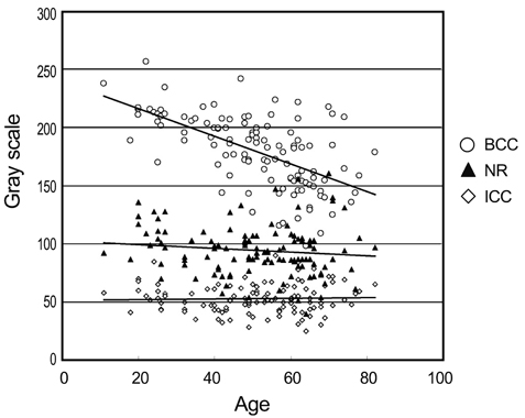
Fig. 2 Correlation between the gray scales and age. BCC = brightest cupping center; NR = nasal rim; ICC = interclass correlation coefficient.
Reference
-
1. Pederson JE, Anderson DR. The mode of progressive disc cupping in ocular hypertension and glaucoma. Arch Ophthalmol. 1980. 98:490–495.2. Nagin P, Schwartz B. Detection of increased pallor over time. Computerized image analysis in untreated ocular hypertension. Ophthalmology. 1985. 92:252–261.3. Gloster J. The colour of the optic disc. Doc Ophthalmol. 1969. 26:155–163.4. Miller JM, Caprioli J. Videographic quantification of optic disc pallor. Invest Ophthalmol Vis Sci. 1988. 29:320–323.5. Vilser W, Nagel E, Seifert BU, et al. Quantitative assessment of optic nerve head pallor. Physiol Meas. 2008. 29:451–457.6. Bearer EL. Overview of image analysis, image importing, and image processing using freeware. Curr Protoc Mol Biol. 2003. Chapter 14:Unit 14.15.7. Schwartz B, Kern J. Scanning microdensitometry of optic disc pallor in glaucoma. Arch Ophthalmol. 1977. 95:2159–2165.8. Jonas JB, Budde WM. Diagnosis and pathogenesis of glaucomatous optic neuropathy: morphological aspects. Prog Retin Eye Res. 2000. 19:1–40.9. Rankin SJ, Drance SM. Peripapillary focal retinal arteriolar narrowing in open angle glaucoma. J Glaucoma. 1996. 5:22–28.10. Albon J, Farrant S, Akhtar S, et al. Connective tissue structure of the tree shrew optic nerve and associated ageing changes. Invest Ophthalmol Vis Sci. 2007. 48:2134–2144.11. Varma R, Tielsch JM, Quigley HA, et al. Race-, age-, gender-, and refractive error-related differences in the normal optic disc. Arch Ophthalmol. 1994. 112:1068–1076.
- Full Text Links
-
- Actions
-
Cited
- CITED
-
- Close
- Share
-
- Similar articles
-
- Letter to the Editor: Quantitative Analysis of Optic Disc Color
- Comparison of Optic Disc Appearance in Anterior ischemic optic neuropathy and Optic neuritis
- On the Ratio to the Physiologic Cupping and Optic Disc. and Optic Disc
- The Effect of Optic Disc Size or Age on Evaluation of Optic Disc Parameters
- Measurements of the Diameter and Area of the Optic Disc
