Hibernoma in Psoas Muscle: A Case Report
- Affiliations
-
- 1Department of Orthopedic Surgery, Yonsei University College of Medicine, Korea. mes1007@yumc.yonsei.ac.kr
- 2Department of Orthopaedic Surgery, National Health Insurance Corporation Ilsan Hospital, Korea.
- 3Department of Diagnostic Pathology, Yonsei University College of Medicine, Korea.
Abstract
- Hibernoma is a rare benign tumor of a brown fat origin with hypervascularity. Although the magnetic resonance imaging features resemble a liposarcoma, its malignant potential is unknown. A complete local excision with meticulous hemostasis is the treatment of choice. We present a case of hibernoma in the psoas muscle with a review of the relevant literature.
Keyword
MeSH Terms
Figure
-
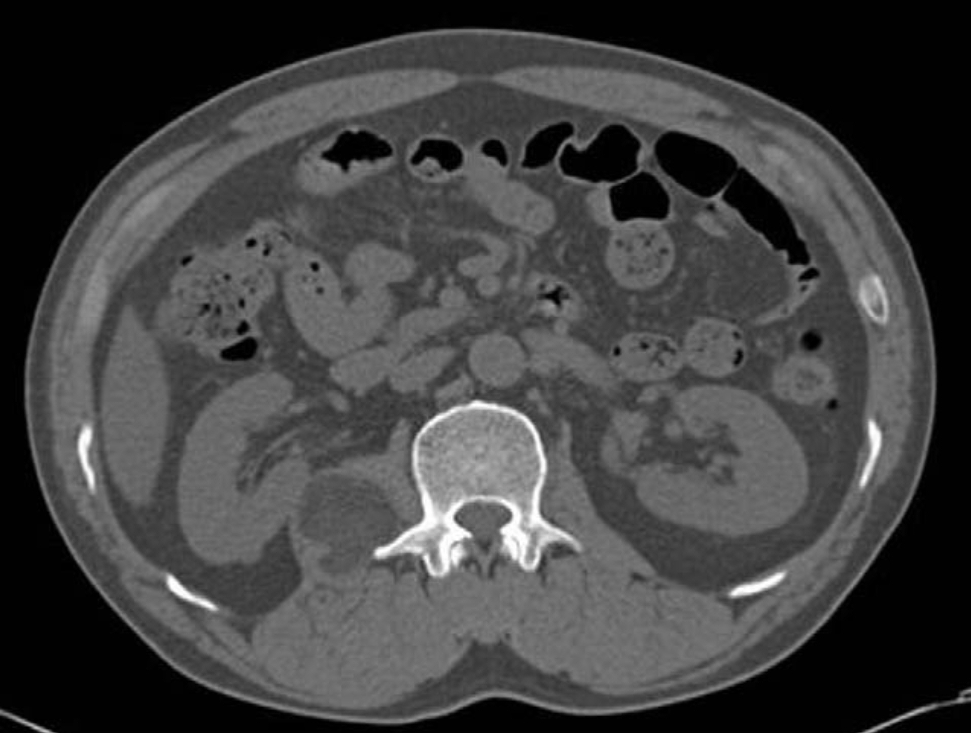
Fig. 1. On computed tomography, about 4 × 4 cm sized, lobu-ated and multiseptated fatty mass was noted in right psoas muscle area extending over T12 to L2 vertebra. The mass showed an increased attenuation compared with subcutaneous fat, and decreased attenuation compared with muscle.
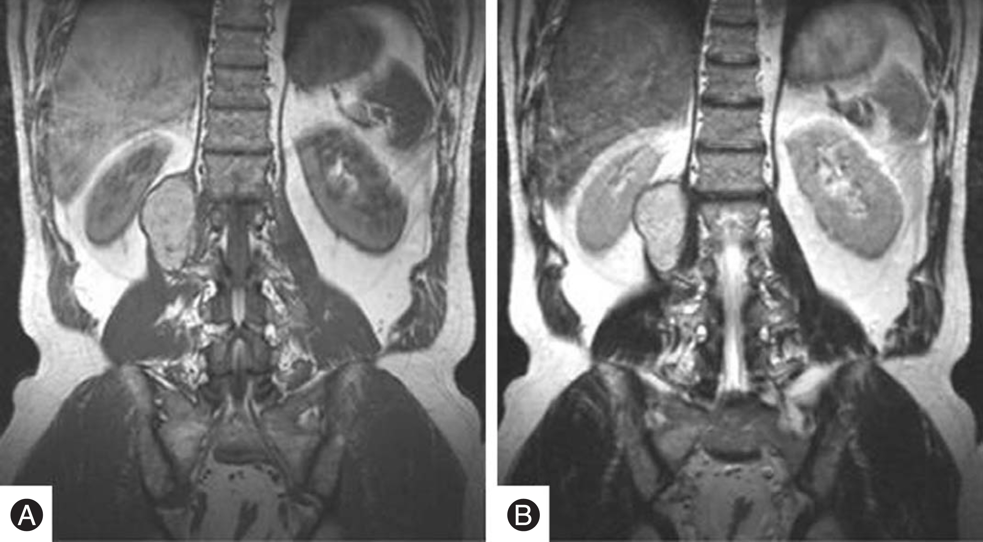
Fig. 2. On preoperative MR imaging, a well marginated and lobulated mass was found on posterior aspect of right kidney being contiguous to right psoas muscle. (A) On T1 weighted imaging, the signal intensity of the mass was intermediate between those of skeletal muscle and subcutaneous fat. (B) On T2 weighted imaging, the mass was heterogenous with somewhat increased signal intensity.
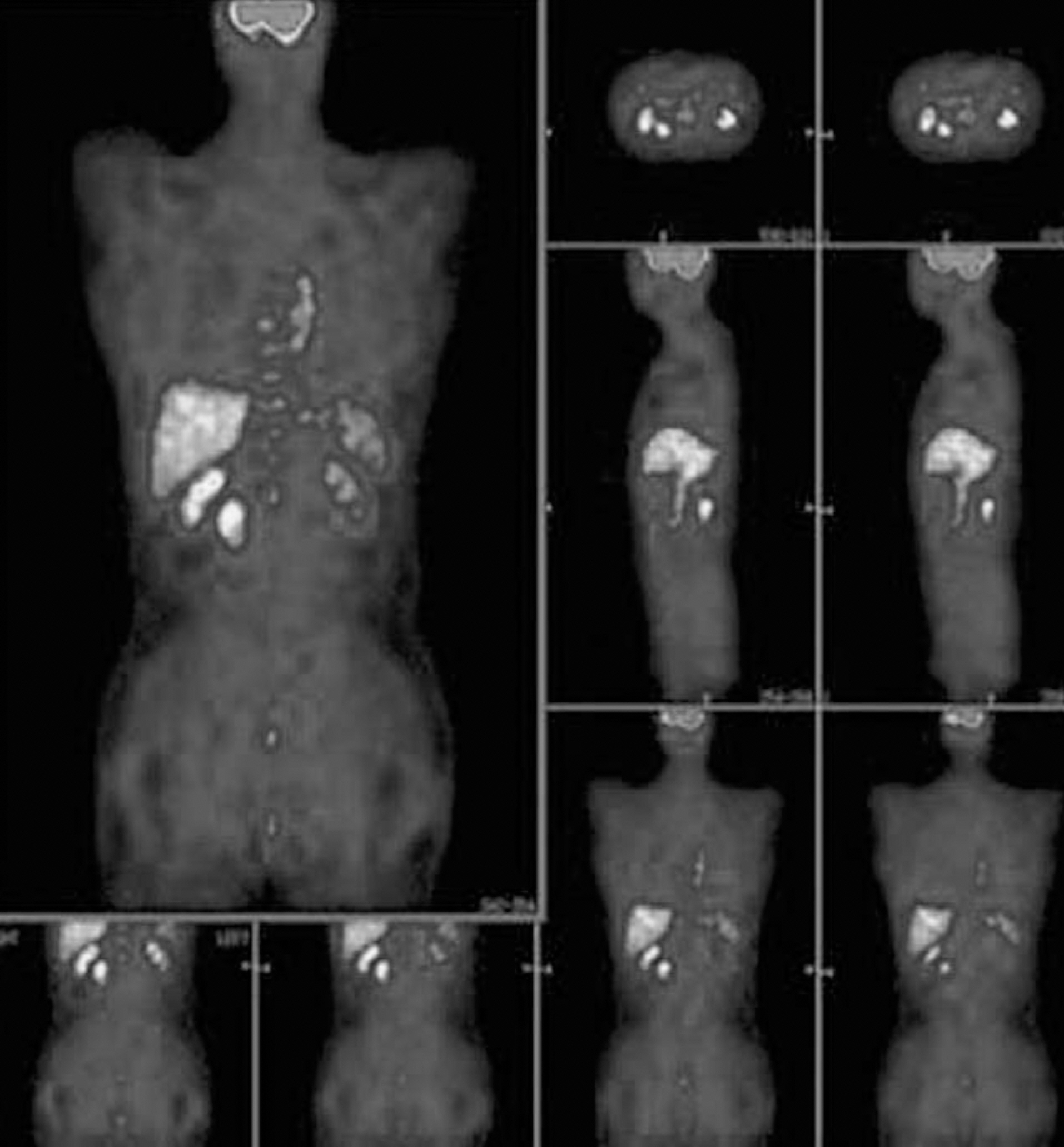
Fig. 3. On PET scan, there was significantly increased FDG uptake on the mass. However there was no increased uptake in other parts of the body indicating metastasis.
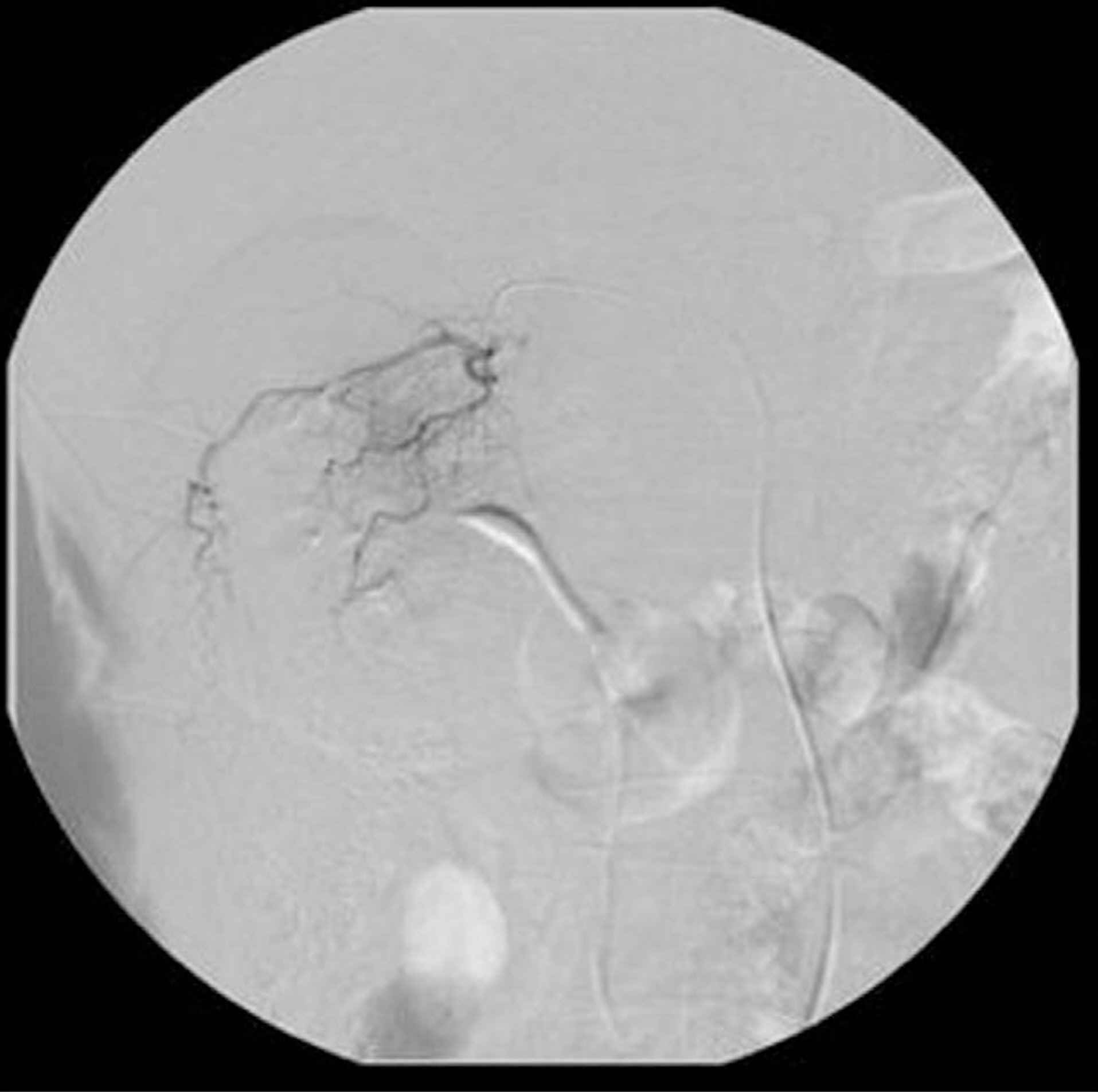
Fig. 4. Preoperative angiography showed a hypervascular mass being fed from branch vessels of 1-2, 2-3, 3-4 lumbar artery. Preoperative embolization was performed for the prevention of intraoperative massive bleeing and extra-compartmental contamination in case of malignancy.
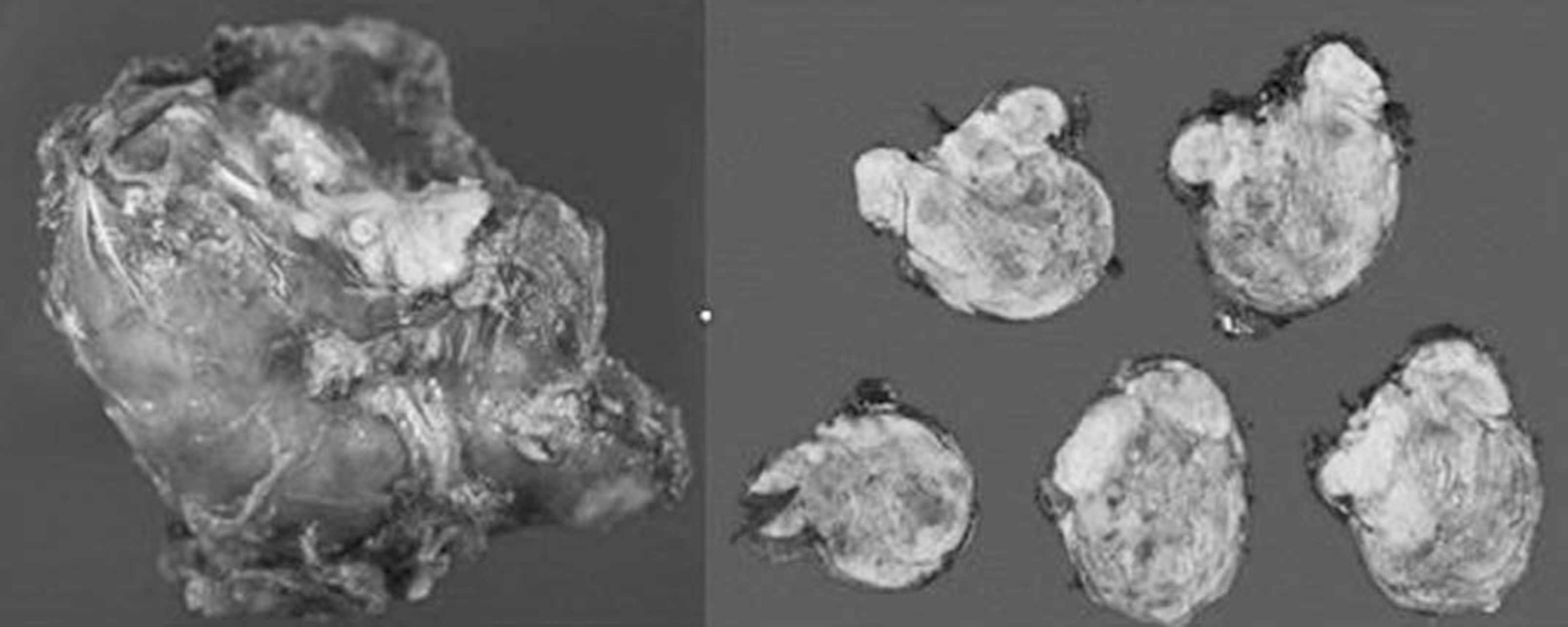
Fig. 5. On gross examination, the mass was about 7 × 4.5 × 4 cm sized and encapsulated with rubbery hard consistency. On serical sections, the cut surface showed multi-lobulation with intervening streaks.
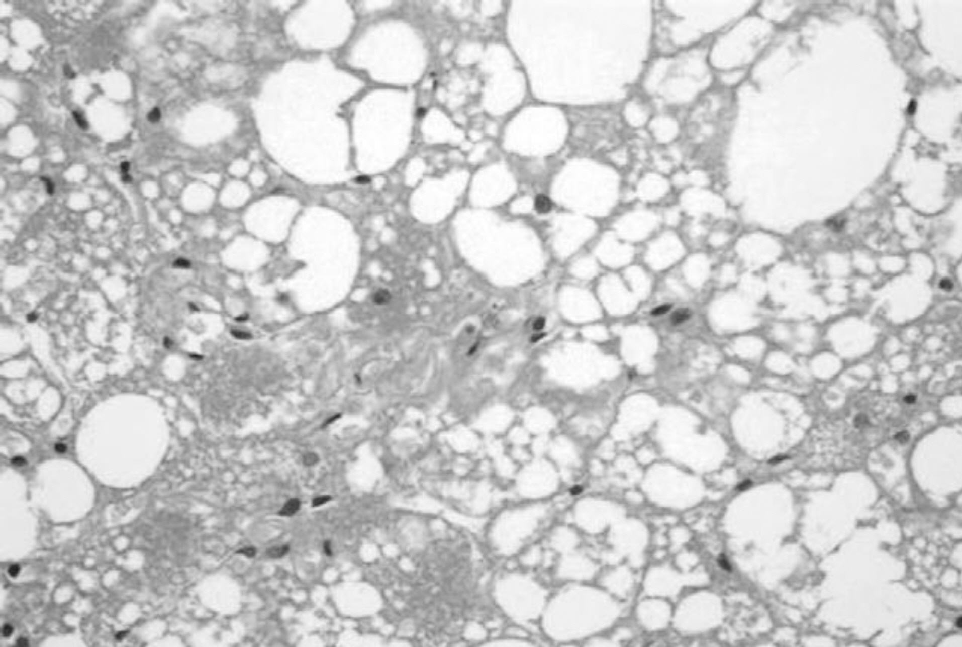
Fig. 6. Histologically, the mass showed a mixture of brown and white adipose cells and was divided into lobules by well vascularized connective tissue. The brown adipose cells are characterized by polygonal, multivacuolated cells with abundant, granular cytoplasm and small, central nucleus.(H-E stain, x100)
Reference
-
1). Meckel H. On a Pseudolipoma of the breast. Beitr Pathol Anat. 1906; 39:152–157.2). Furlong MA, Fanburg-Smith JC, Miettinen M. The Morphologic Spectrum of Hibernoma: A Clinicopatholog-ic Study of 170 Cases. The American Journal of Surgical Pathology. 2001; 25:809–814.3). Enzinger FM, Weiss SW. Soft Tissue Tumors. London: CV Mosby;p. 234–241. 1983.4). Lee TJ, Park IS, Kim MG, Cho KJ, Moon KH, Lim KY. Axillary Hibernoma: MRI Characteristics. J of Korean Orthop Assoc. 2004; 39:335–338.5). Lewandowski PJ, Weiner SD. Hibernoma of the Medial Thigh. Clinical Orthopaedics and related research. 1996; 330:198–201.
Article6). Cook MA, Stern M, de Silva RD. Case report: MRI of hibernoma. J Comput Assis Tomogr. 1996; 20:333–335.7). McLane RC, Meyer LC. Axillary Hibernoma: Review of the literature with report of a case examined angiographi-cally. Radiology. 1978; 127:673–679.
Article8). Munk PL, Lee MJ, Janzen DL. Lipoma and liposarcoma: Evaluation using CT and MR imaging. AHR AM J Roentgenol. 1997; 169:589–594.
Article
