Gangliocytoma Mimicking Extra-axial Tumor: A Report of Two Cases
- KMID: 754113
- DOI: http://doi.org/10.3348/kjr.2001.2.2.108
Abstract
- We report two cases of supratentorial gangliocytomas mimicking an extra-axial tumor. MR imaging indicated that the tumors were extra-axial, and meningiomas were thus initially diagnosed. Relative to gray matter, the tumors were hypointense on T1-weighted images and hyperintense on T2-weighted images. On contrast-enhanced T1-weighted images, homogeneous enhancement was observed, while CT scanning revealed calcification in one of the two cases.
Keyword
MeSH Terms
Figure
-
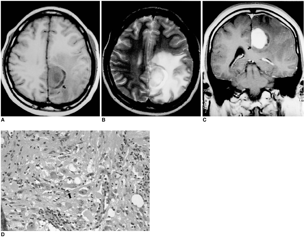
Fig. 1 (Case 1) A 22-year-old woman who complained of right hemiparesis and hemiparesthesia. A. Axial T1-weighted MR image shows a well demarcated tumor in the left para-midline area, with peritumoral edema. The tumor is of slightly low signal intensity and has a broad dural base on the falx cerebri and crescentic peritumoral cysts (arrow). B. Axial T2-weighted MR image shows a high signal intensity tumor with marked peritumoral edema. C. Gadolinium-enhanced coronal T1-weighted MR image demonstrates strong homogeneous enhancement, with the dural tail (arrow) attached to the falx cerebri. D. Microscopic examination indicates that the tumor is composed of ganglion cells (arrow) that contain prominent and eccentric nucleoli, and abundant bluish cytoplasm (hematoxylin-eosin, original magnification ×200).

Fig. 2 (Case 2) A 57-year-old woman who complained of headache and dizziness. A. Axial T1-weighted MR image shows a small cortical tumor of slightly low signal intensity and with an exophytic component (arrow). Note the presence of subdural hygroma adjacent to the tumor (open arrows). B. Axial T2-weighted MR image reveals high signal intensity (arrow). Discrimination between the exophytic extra-axial component and surrounding cerebrospinal fluid is difficult. C. Gadolinium-enhanced T1-weighted sagittal image shows that the tumor is located in the cortex, has an exophytic growth pattern (arrows), and shows homogeneous enhancement. D. Unenhanced CT scan reveals tumor calcification (arrow). E. Microscopic examination (low-power field) shows that the tumor is attached to the dura mater (arrow). (magnification ×40; hematoxylin-eosin stain). F. Immunohistochemical staining demonstrates positive staining of the cytoplasm and process of some ganglion cells with synaptophysin (arrows).
Reference
-
1. Zulch KJ. Brain Tumours: Their Biology and Pathology. 1986. Berlin: Springer-Verlag;184.2. Furie DM, Felsberg GJ, Tien RD, et al. MRI of gangliocytoma of the cerebellum and spinal cord. J Comput Assist Tomogr. 1993. 17(3):488–491.3. Altman NR. MR and CT characteristics of gangliocytoma, a rare cause of epilepsy in children. AJNR. 1988. 9:917–921.4. Sherazi ZA. Gangliocytoma: magnetic resonance imaging characteristics. Singapore Med J. 1998. 39(8):373–375.5. Izukawa D, Lach B, Benoit B. Gangliocytoma of the cerebellum: ultrastructure and immunohistochemistry. Neurosurgery. 1988. 22:576–581.6. Kawamoto K, Yamanouchi Y, Suwa J, Kurimoto T, Matsumura H. Ultrastructural study of a cerebral gangliocytoma. Surg Neurol. 1985. 24:541–549.7. Peretti-Viton P, Perez-Castillo AM, Raybaud C, et al. Magnetic resonance imaging in gangliogliomas and gangliocytomas of the nervous system. J Neuroradiol. 1991. 18(2):189–199.8. Itoh Y, Yagishita S, Chiba Y. Cerebral gangliocytoma. An ultrastructural study. Acta Neuropathol. 1987. 74:169–178.
- Full Text Links
-
- Actions
-
Cited
- CITED
-
- Close
- Share
-
- Similar articles
-
- A Case of Acromegaly Caused by Mixed Gangliocytoma-Adenoma of the Pituitary Gland
- Supratentorial Anaplastic Ependymoma Mimicking an Extra-Axial Tumor: A Case Report
- Extra-Axial Medulloblastoma in the Cerebellar Hemisphere
- A Cerebral Astroblastoma Mimicking an Extra-axial Neoplasm
- A Rare Case of Concomitant Intramedullary Gangliocytoma at the Cervicomedullary Junction in Patient with Neuroendocrine Tumor of Lung
