Ultrasound Evaluation of Normal and Abnormal Fetuses: Comparison of Conventional, Tissue Harmonic, and Pulse-Inversion Harmonic Imaging Techniques
- Affiliations
-
- 1Department of Radiology, Samsung Medical Center, Sungkyunkwan University School of Medicine. bhkim@smc.samsung.co.kr
- 2Department of Diagnostic Radiology, Bundang CHA General Hospital, Pochon CHA University College of Medicine.
- 3Department of Radiology, Pundang Je-saeng General Hospital.
- 4Department of Obstetrics and Gynecology, Samsung Medical Center, Sungkyunkwan University School of Medicine.
- 5Department of Radiology, Miz Medi Hospital.
- 6Department of Preventive Medicine, College of Medicine, Korea University.
- KMID: 754023
- DOI: http://doi.org/10.3348/kjr.2003.4.3.184
Abstract
OBJECTIVE
To determine the usefulness of tissue harmonic imaging (THI) and pulse-inversion harmonic imaging (PIHI) in the evaluation of normal and abnormal fetuses. MATERIALS AND METHODS: Forty-one pregnant women who bore a total of 31 normal and ten abnormal fetuses underwent conventional ultrasonography (CUS), and then THI and PIHI. US images of six organ systems, namely the brain, spine, heart, abdomen, extremities and face were compared between the three techniques in terms of overall conspicuity and the definition of borders and internal structures. RESULTS: For the brain, heart, abdomen and face, overall conspicuity at THI and PIHI was significantly better than at CUS (p < 0.05). There was, though, no significant difference between THI and PIHI. Affected organs in abnormal fetuses were more clearly depicted at THI and PIHI than at CUS. CONCLUSION: Both THI and PIHI appear to be superior to CUS for the evaluation of normal or abnormal structures, particularly the brain, heart, abdomen and face.
Figure
-
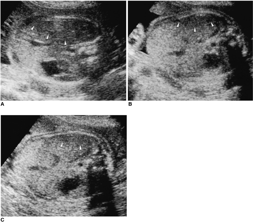
Fig. 1 Transverse views of fetal abdomen: (A) CUS, (B) THI, (C) PIHI. Soft-tissue contrast is better at B and C than at A. Artifactual hypoechoic areas in the right lobe of the liver near the transducer (arrowheads) are smaller at B and C than at A, and smallest at C.
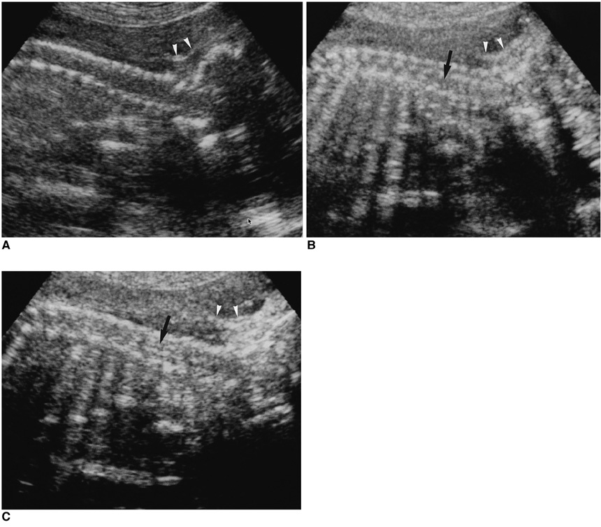
Fig. 2 Longitudinal scans of fetal cervical spine: (A) CUS, (B) THI, (C) PIHI. The margins of bony structures are clearer at A, since the posterior sonic shadowing of the individual bones is less prominent. Soft tissue contrast, however, seems to be higher at B and C. Layers of suboccipital scalp (arrowheads) are clearly demonstrated at B, and the spinal cord (arrows) is clearly visible at C.
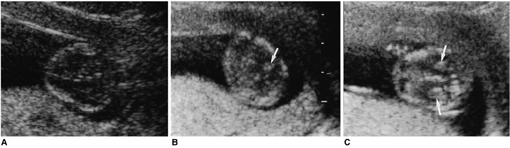
Fig. 3 Transverse scan of the fetal head at 12 weeks' gestation: (A) CUS, (B) THI, (C) PIHI. The choroid plexuses (arrows) present in lateral ventricles are clearly visible at C, but internal structures are not clearly depicted at A.
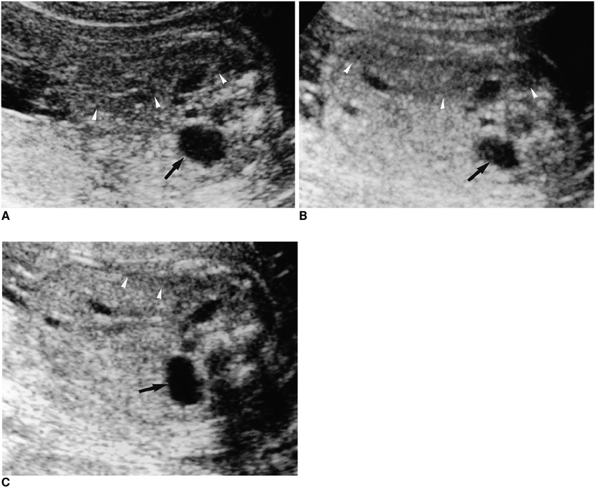
Fig. 4 Transverse scan through fetal kidneys: (A) CUS, (B) THI, (C) PIHI. The dilated renal pelvis (arrows) is clearly demonstrated by all three techniques, though soft tissue contrast is clearest at PIHI. Note the presence of the smallest near-field artifact (arrowheads) at PIHI.
Reference
-
1. Tranquart F, Grenier N, Eder V, et al. Clinical use of ultrasound tissue harmonic imaging. Ultrasound Med Biol. 1999. 25:889–894.2. Caidahl K, Kazzam E, Lidberg J, et al. New concept in echocardiography: harmonic imaging of tissue without use of contrast agent. Lancet. 1998. 352:1264–1270.3. Lindner JR, Villanueva FS, Dent JM, et al. Assessment of resting perfusion with myocardial contrast echocardiography: theoretical and practical considerations. Am Heart J. 2000. 139:231–240.4. Spencer KT, Bednarz J, Rafter PG, et al. Use of harmonic imaging without echocardiographic contrast to improve two-dimensional image quality. Am J Cardiol. 1998. 82:794–799.5. Schwarz KQ, Chen X, Steinmetz S, et al. Harmonic imaging with Levovist. J Am Soc Echocardiogr. 1997. 10:1–10.6. Postert T, Federlein J, Weber S, et al. Second harmonic imaging in acute middle cerebral artery infarction: preliminary results. Stroke. 1999. 30:1702–1706.7. Seidel G, Greis C, Sonne J, et al. Harmonic gray scale imaging of the human brain. J Neuroimaging. 1999. 9:171–174.8. Hann LE, Bach AM, Cramer LD, et al. Hepatic sonography: comparison of tissue harmonic and standard sonography techniques. AJR Am J Roentgenol. 1999. 173:201–206.9. Kono Y, Moriyasu F, Nada T, et al. Gray scale second harmonic imaging of the liver: a preliminary animal study. Ultrasound Med Biol. 1997. 23:719–726.10. Jang HJ, Lim HK, Lee WJ, Kim SH, Kim KA, Kim EY. Ultrasonographic evaluation of focal hepatic lesions: comparison of pulse-inversion harmonic, tissue harmonic, and conventional imaging techniques. J Ultrasound Med. 2000. 19:293–299.11. Shapiro RS, Wagreich J, Parsons RB, et al. Tissue harmonic imaging sonography: evaluation of image quality compared with conventional sonography. AJR Am J Roentgenol. 1998. 171:1203–1206.12. Desser TS, Jeffrey RB Jr, Lane MJ, Ralls PW. Tissue harmonic imaging: utility in abdominal and pelvic sonography. J Clin Ultrasound. 1999. 27:135–142.13. Sehgal CM, Arger PH, Pugh CR, Kirchofer JI, Kotlar EY, Bovee KC. Comparison of power Doppler and B-scan sonography for renal imaging using a sonographic agent. J Ultrasound Med. 1998. 17:751–756.14. Claudon M, Barnewolt CE, Taylor GA, et al. Renal blood flow in pigs: changes depicted with contrast-enhanced harmonic US imaging during acute urinary obstruction. Radiology. 1999. 212:725–731.15. Taylor GA, Barnewolt CE, Claudon M, et al. Depiction of renal perfusion defects with contrast-enhanced harmonic sonography in a porcine model. AJR Am J Roentgenol. 1999. 173:757–760.16. Schmiedl UP, Carter S, Martin RW, et al. Sonographic detection of acute parenchymal injury in an experimental porcine model of renal hemorrhage: gray-scale imaging using sonographic contrast agent. AJR Am J Roentgenol. 1999. 173:1289–1294.17. Kim B, Lim HK, Choi MH, et al. Detection of parenchymal abnormalities in acute pyelonephritis by pulse-inversion harmonic imaging with or without microbubble US contrast agent: correlation with CT. J Ultrasound Med. 2001. 20:5–14.18. Callen PW. Callen PW, editor. The obstetric ultrasonographic examination. Ultrasonography in obstetrics and gynecology. 2000. 4th ed. Philadelphia, Pensylvania, U.S.A.: W. B. Saunders Company;1–16.19. Lee SH, Cho JY, Song MJ, et al. Prenatal ultrasound findings of fetal neoplasms. Korean J Radiol. 2002. 3:64–73.20. Ozkutlu S, Saraclar M. The accuracy of antenatal fetal echocardiography. Turk J Pediatr. 1999. 41:349–352.21. Barnhard Y, Bar-Hara I, Divon MY. Accuracy of intrapartum estimates of fetal weight: effect of oligohydramnios. J Reprod Med. 1996. 41:907–910.22. Sivit CJ, Hill MC, Larsen JW, et al. The sonographic evaluation of fetal anomalies in oligohydramnios between 16 and 30 weeks gestation. AJR Am J Roentgenol. 1986. 146:1277–1281.23. Cohen J. A coefficient of agreement for nominal scales. Educational Psychological Measurement. 1960. 20:37–39.24. Powers J, Averkiou M, Hope-Simpson D. Contrast imaging methods. 1998. In : Proceedings of the First Annual Symposium on Ultrasound Contrast for Radiological Diagnosis; 2-3 October; Toronto, Canada. –18.25. Senior R, Soman P, Khattar RS, et al. Improved endocardial visualization with second harmonic imaging compared with fundamental two-dimensional echocardiographic imaging. Am Heart J. 1999. 138:163–168.26. Bauer A, Hauff P, Lazenby J, et al. Wideband harmonic imaging: a novel contrast ultrasound imaging technique. Eur Radiol. 1999. 9:Suppl 3. S364–S367.27. Egerton IB, Vella G, Barnett S. Experimental test of harmonic contribution to the thermal index for soft tissue. J Ultrasound Med. 1999. 18:81–86.
- Full Text Links
-
- Actions
-
Cited
- CITED
-
- Close
- Share
-
- Similar articles
-
- Comparison Study among Conventional, Tissue Harmonic and Pulse Inversion Harmonic Images to Evaluate Pleural Effusion and Ascites
- Ultrasonographic Evaluation of the Normal Kidney: Comparison of Fundamental, Tissue Harmonic, and Pulse Inversion Harmonic Imaging
- Sonographic Evaluation of Breast Nodules: Comparison of Conventional, Real-Time Compound, and Pulse-Inversion Harmonic Images
- Pulse Inversion Harmonic Imaging and Stimulated Acoustic Emission of Hepatic Masses
- Hepatic Hemangiomas: Spectrum of US Appearances on Gray-scale, Power Doppler, and Contrast-Enhanced US
