Pediatric Central Nervous System Vascular Malformation : Pathological Review with Diagram
- Affiliations
-
- 1Department of Pathology, Severance Hospital, Yonsei University College of Medicine, Seoul, Korea
- KMID: 2554863
- DOI: http://doi.org/10.3340/jkns.2024.0006
Abstract
- Pediatric central nervous system (CNS) vascular malformations are a group of abnormal blood vessel formations within the brain or spinal cord in children. The most crucial point of pediatric CNS vascular malformation is that no golden standard classifications exist. In addition, there is a big gap in knowledge and the viewpoint of clinicians, radiologists, and pathologists. In addition, many genes associated with pediatric CNS vascular malformation, such as Sturge-Weber-Dimitri syndrome with guanine nucleotide-binding protein G(q) subunit alpha (GNAQ) gene mutation, and cavernous malformations with cerebral cavernous malformations 1 (CCM1), CCM2, and CCM3 gene mutation, were recently revealed. For proper therapeutic approaches, we must understand the lesions’ characterizations in anatomical, morphological, and functional views. In this review, the author would like to provide basic pediatric CNS vascular malformation concepts with understandable diagrams. Thus, the author hopes that it might be helpful for the proper diagnosis and treatment of CNS pediatric vascular malformations.
Figure
-
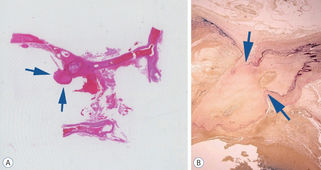
Fig. 1. The balloon-like protruding vascular lesion with hemorrhage (arrows) is noted (Hematoxylin and Eosin, ×1). B : The elastic Van Gieson’s (EVG) staining shows abrupt loss of elastic fibers (black colors) (arrows) in the aneurysmal lesion (EVG, ×12).
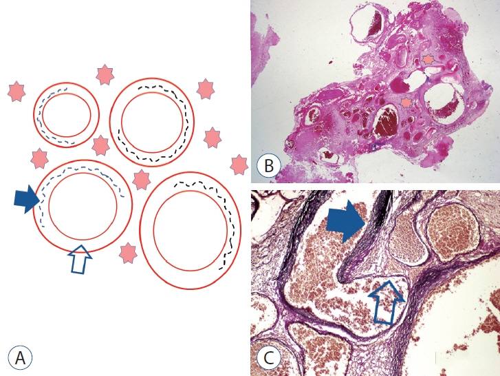
Fig. 2. A : The diagram of arteriovenous malformation (AVM). Variable-sized arterialized vessels with incomplete elastic fibers (black line) are seen. The gliotic tissues (pink stars) are in between the vessels and the surrounding lesion. The blue solid arrow indicates the presence of elastic fibers, and the open arrow indicates the absence of elastic fibers. B : The microscopic findings of AVM (Hematoxylin and Eosin, ×12). The intervening gliotic tissues (pink stars) are noted. C : The elastic Van-Gieson staining (EVG, ×100). An arterialized vein shows an incomplete elastic layer (blue solid arrow : presence; open arrow : absence).
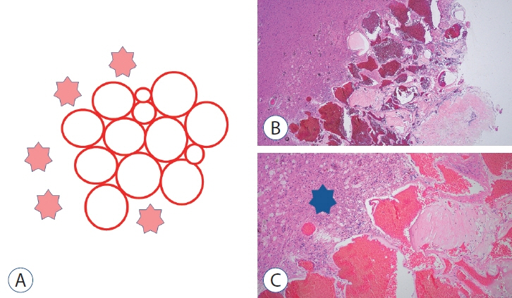
Fig. 3. A : The diagram of cavernous malformations (CMs). Hyalinized vessels are located closely without any intervening brain parenchyma. Surrounding the lesion, the gliotic tissues (pink stars) are noted. B : The microscopic findings of CM (Hematoxylin and Eosin [H&E], ×40). The hyalinized vessels are closed without intervening gliotic tissues. C : Surrounding the lesion, there are extensive hemosiderin-laden macrophages and reactive gliosis. (blue star) (H&E, ×100).
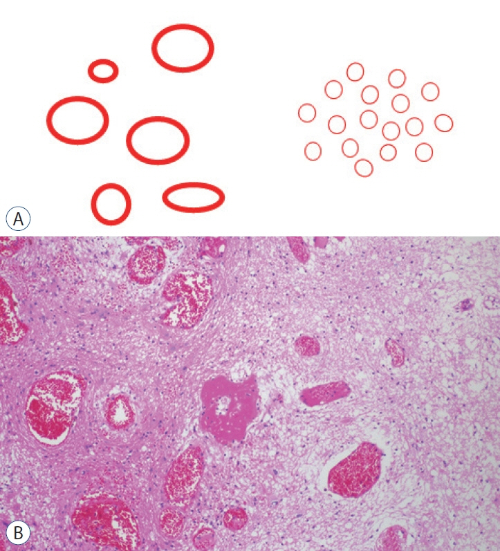
Fig. 4. A : The diagrams of developmental venous anomalies (DVA) (left) and capillary telangiectasias (right). There are dilated vessels with intervening normal blood parenchyma. The calibers of DVA between capillary telangiectasia are different. B : A typical histological finding of DVA (Hematoxylin and Eosin, ×100).
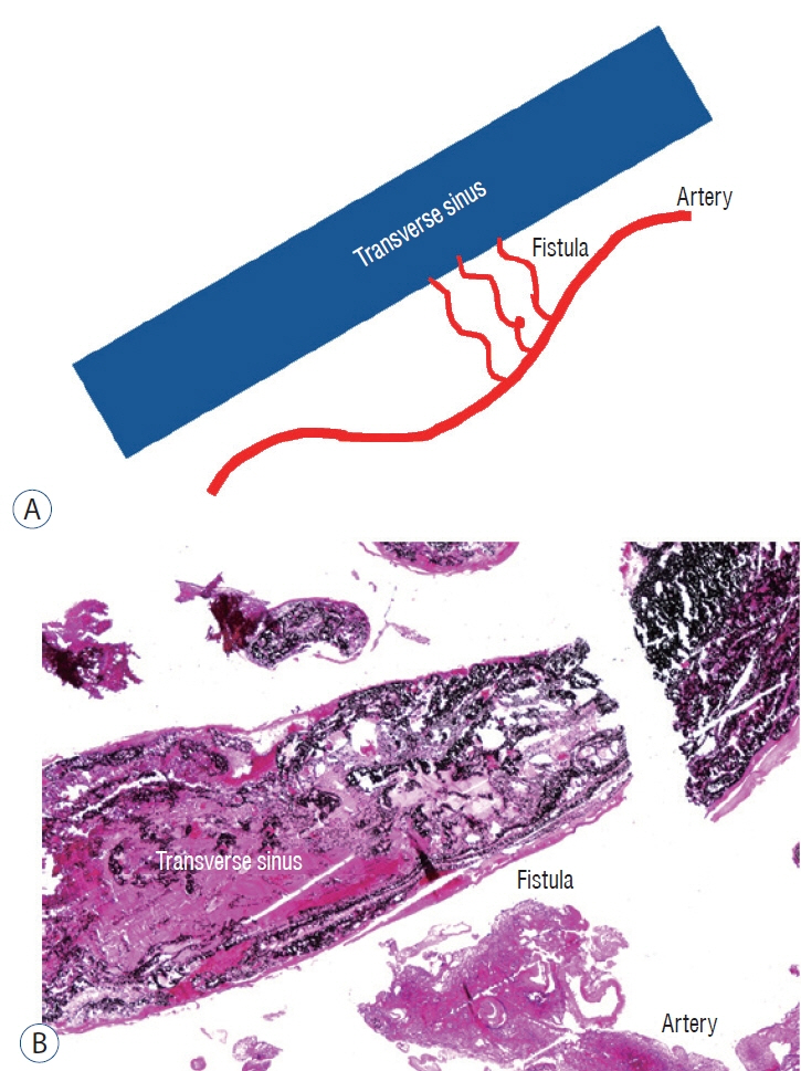
Fig. 5. A : The diagram of dural arteriovenous fistulas. In dura, there are abnormal connections between arteries and veins (transverse sinus). B : The embolized transverse sinus with arteries and fistulas is noted (Hematoxylin and Eosin, ×12).
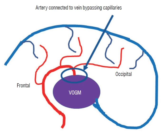
Fig. 6. The diagram of vein of Galen malformations (VOGM).
Reference
-
References
1. Ahlawat S, Fayad LM, Durand DJ, Puttgen K, Tekes A. International society for the study of vascular anomalies classification of soft tissue vascular anomalies: survey-based assessment of musculoskeletal radiologists’ use in clinical practice. Curr Probl Diagn Radiol. 48:10–16. 2019.
Article2. Beez T, Steiger HJ, Hänggi D. Evolution of management of intracranial aneurysms in children: a systematic review of the modern literature. J Child Neurol. 31:773–783. 2016.
Article3. Castillo-Rangel C, Marín G, Hernandez-Contreras KA, Zarate-Calderon C, Vichi-Ramirez MM, Cortez-Saldias W, et al. Atlas of nervous system vascular malformations: a systematic review. Life (Basel). 12:1199. 2022.
Article4. Krings T, Geibprasert S, terBrugge KG. Pathomechanisms and treatment of pediatric aneurysms. Childs Nerv Syst. 26:1309–1318. 2010.
Article5. Labauge P, Denier C, Bergametti F, Tournier-Lasserve E. Genetics of cavernous angiomas. Lancet Neurol. 6:237–244. 2007.
Article6. Magaki SD, Tashjian R, Vinters HV. Pediatric Vascular Malformations in Gray F, Keohane K (eds). Developmental Neuropathology. ed 2. Hoboken: John Wiley & Sons;2018. p. 251–267.7. Shirley MD, Tang H, Gallione CJ, Baugher JD, Frelin LP, Cohen B, et al. Sturge-Weber syndrome and port-wine stains caused by somatic mutation in GNAQ. N Engl J Med. 368:1971–1979. 2013.
Article
- Full Text Links
-
- Actions
-
Cited
- CITED
-
- Close
- Share
-
- Similar articles
-
- Treatment of pediatric central nervous system infections
- The value of computerized axial tomography of the brain in children with central nervous system disorders
- A Study on CSF Enzyme Activity in Central Nervous System Infections
- A Case of Extradural Cavernous Hemangioma with Reuptured Disc
- Dandy-Walker Malformation Associated with Neurocutaneous Melanosis
