Ewha Med J.
2024 Jan;47(1):e9. 10.12771/emj.2024.e9.
Disseminated cutaneous gout: a rare manifestation of gout
- Affiliations
-
- 1Department of Dermatology, Ewha Womans University College of Medicine, Seoul, Korea
- KMID: 2553301
- DOI: http://doi.org/10.12771/emj.2024.e9
Figure
-
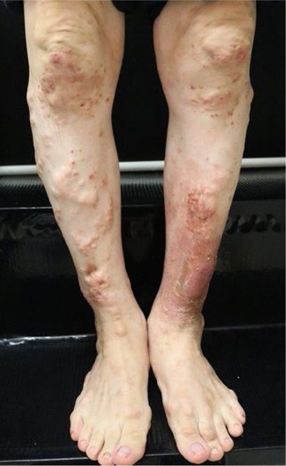
Fig. 1. Multiple yellowish, firm papulonodules had developed on both lower legs. Some lesions showed acute suppuration.
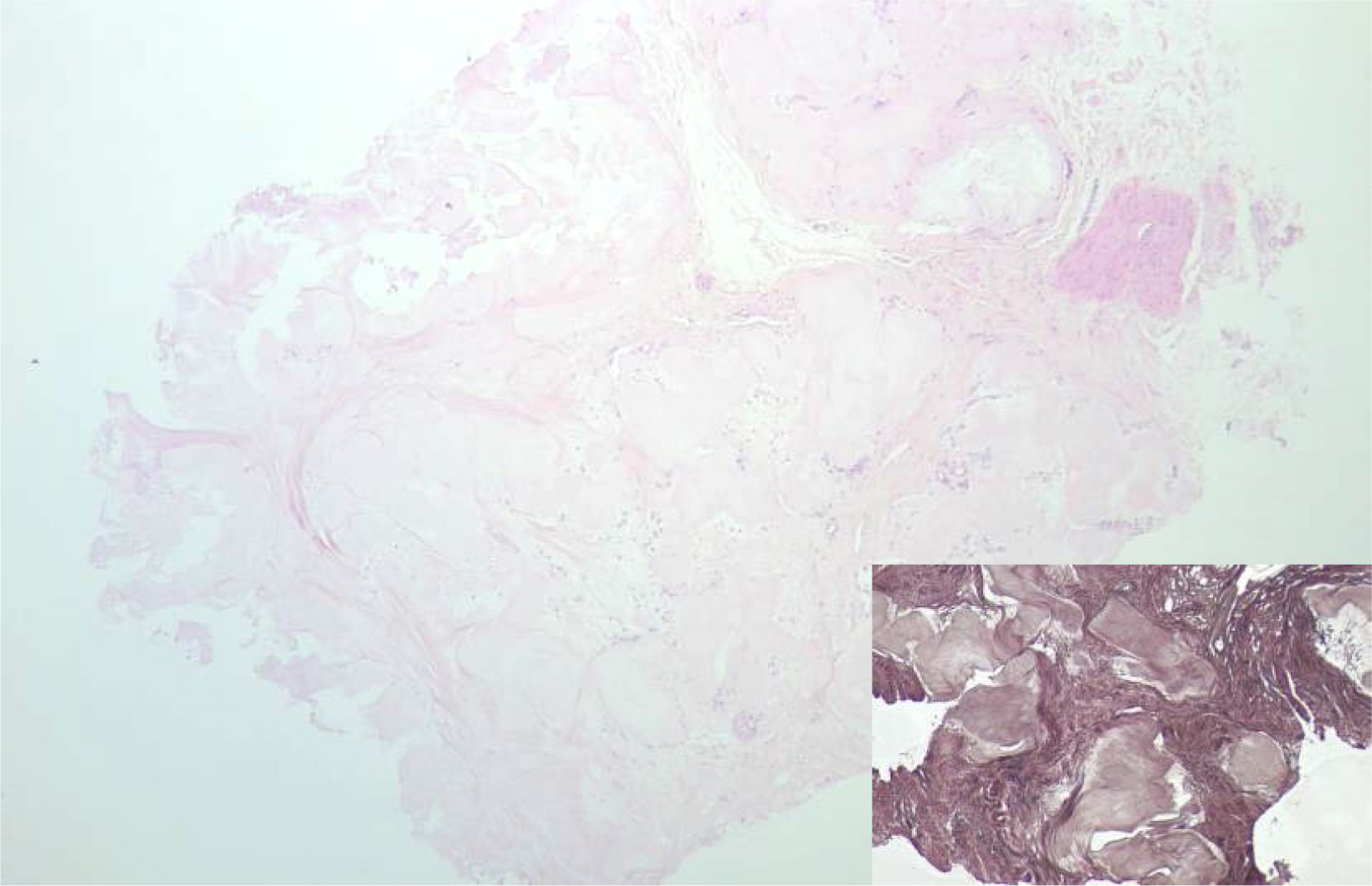
Fig. 2. Well-circumscribed deposition of pinkish amorphous materials was noticed in the deep dermis. Histiocytic infiltration and fibrous reaction were observed surrounding the amorphous materials (hematoxylin and eosin, ×40). Inlet, Von Kossa staining showed no calcium deposition (×100).
Reference
-
References
1. Dalbeth N, Merriman TR, Stamp LK. Gout. Lancet. 2016; 388(10055):2039–2052. DOI: 10.1016/S0140-6736(16)00346-9. PMID: 27112094.
Article2. Fairley JA, Aronson AB. Calcium and other mineral deposition disorders. In. Kang S, Amagai M, Bruckner AL, Enk AH, Margolis DJ, McMichael AJ, Orringer JS, editors. editors. Fitzpatrick’s dermatology. 9th ed. New York: McGraw Hill Education;2019. p. p. 2313–2316.3. Pradhan S, Sinha R, Sharma P, Sinha U. Atypical cutaneous presentation of chronic tophaceous gout: a case report. Indian Dermatol Online J. 2010; 11(2):235–238. DOI: 10.4103/idoj.IDOJ_205_19. PMID: 32477988. PMCID: PMC7247644.
Article4. Guzman R, DeClerck B, Crew A, Peng D, Adler BL. Disseminated cutaneous gout: a rare manifestation of a common disease. Dermatol Online J. 2020; 26(1):4. DOI: 10.5070/D3261047184.
Article
