Maxillary complete denture with posterior zirconia occlusion and mandibular implant support fixed prostheses in completely edentulous patients with orofacial dystonia
- Affiliations
-
- 1Department of Prosthodontics, Dental and Life Sciences Institute, Education and Research Team for Life Science on Dentistry, School of Dentistry, Pusan National University, Yangsan, Republic of Korea
- 2Department of Prosthodontics, Dental Research Institute, Pusan National University Dental Hospital, Yangsan, Republic of Korea
- KMID: 2550433
- DOI: http://doi.org/10.14368/jdras.2023.39.4.237
Abstract
- Orofacial dystonia is a neuromotor disorder that causes irregular or repetitive movements of the face, lips, tongue, and jaw involuntarily, also called tic disorder. Edentulous patients with these symptoms experience functional and aesthetic problems, including difficulty using complete dentures, speech and swallowing difficulties, and orofacial pain. In this case, for a patient with orofacial dystonia who experienced complete edentulism at a relatively young age, restorative treatment was performed with a maxillary complete denture with bilateral posterior zirconia occlusal surfaces and a mandibular implant-supported fixed prosthesis, and continuous smile training was performed. The aim was to improve the aesthetics of facial muscles. As a result of the treatment, the patient was very satisfied with not only improved chewing function and aesthetics, but also regained psychological stability and was able to lead a normal daily life, so we would like to report this.
Keyword
Figure
-
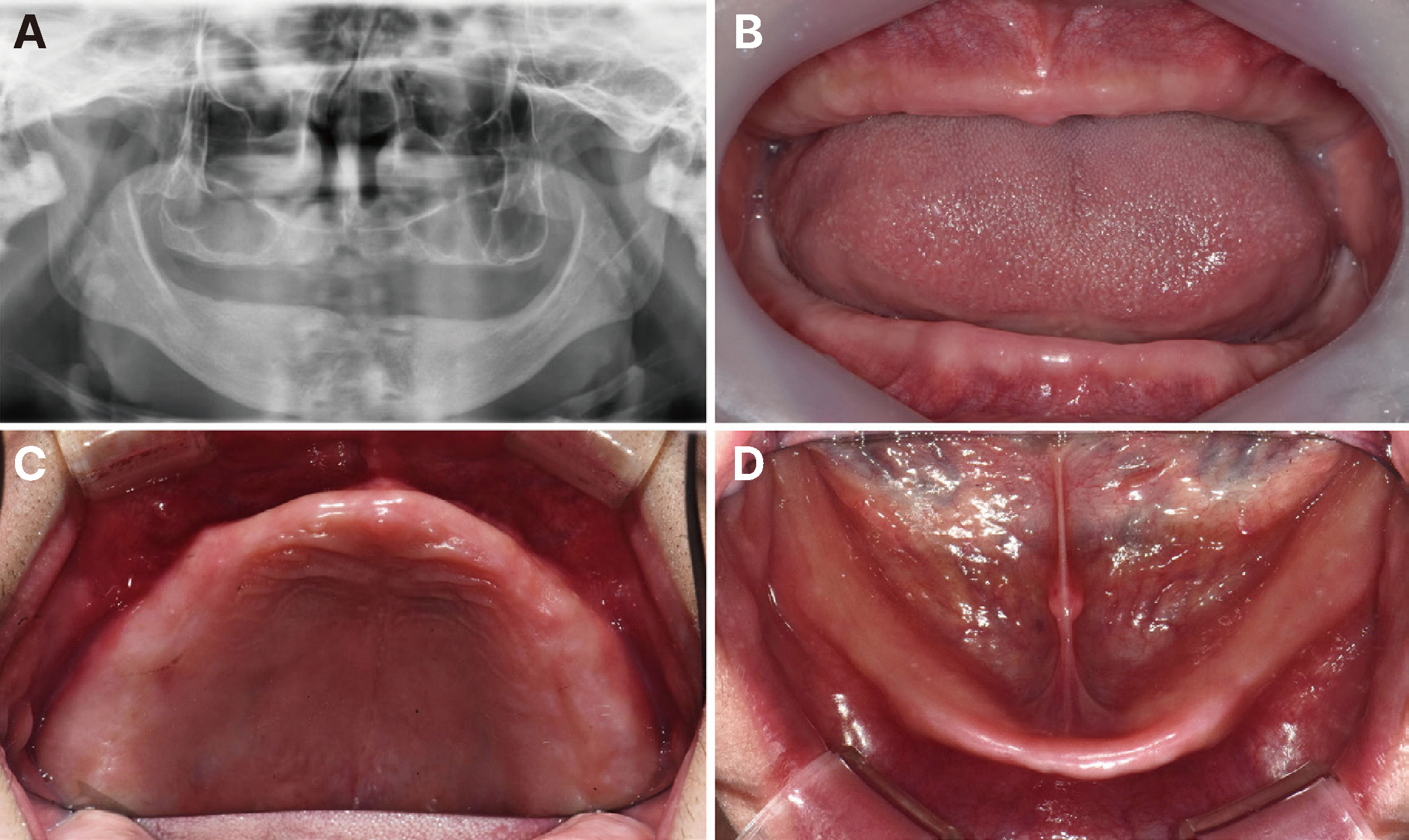
Fig. 1 Panorama and Intraoral view. (A) Initial panoramic radiograph, (B) Frontal view, (C) Maxillary occlusal view, (D) Mandibular occlusal view.
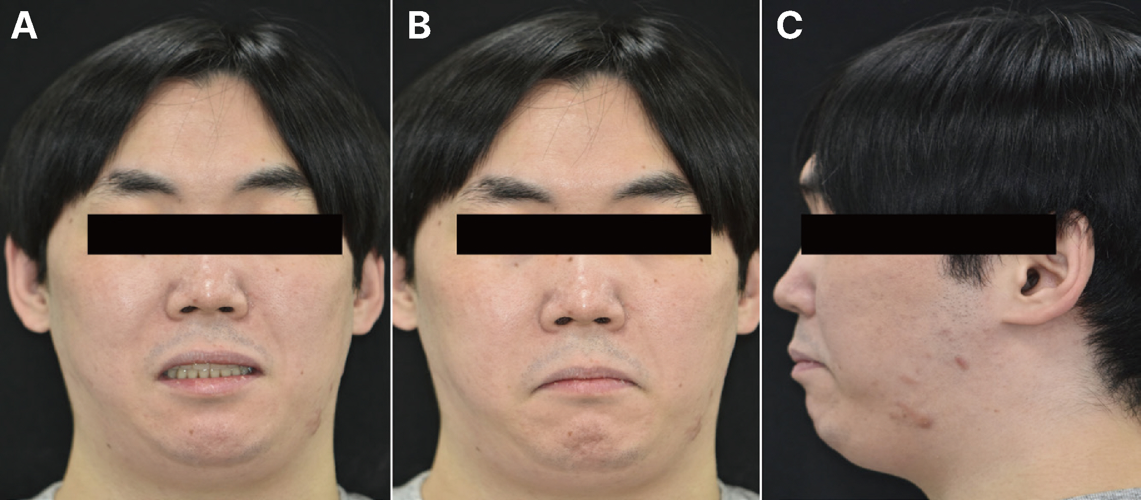
Fig. 2 Extraoral view of patient with Orofacial dystonia. (A) Frontal view with old denture, (B) Frontal view without old denture, (C) Sagittal view without old denture.
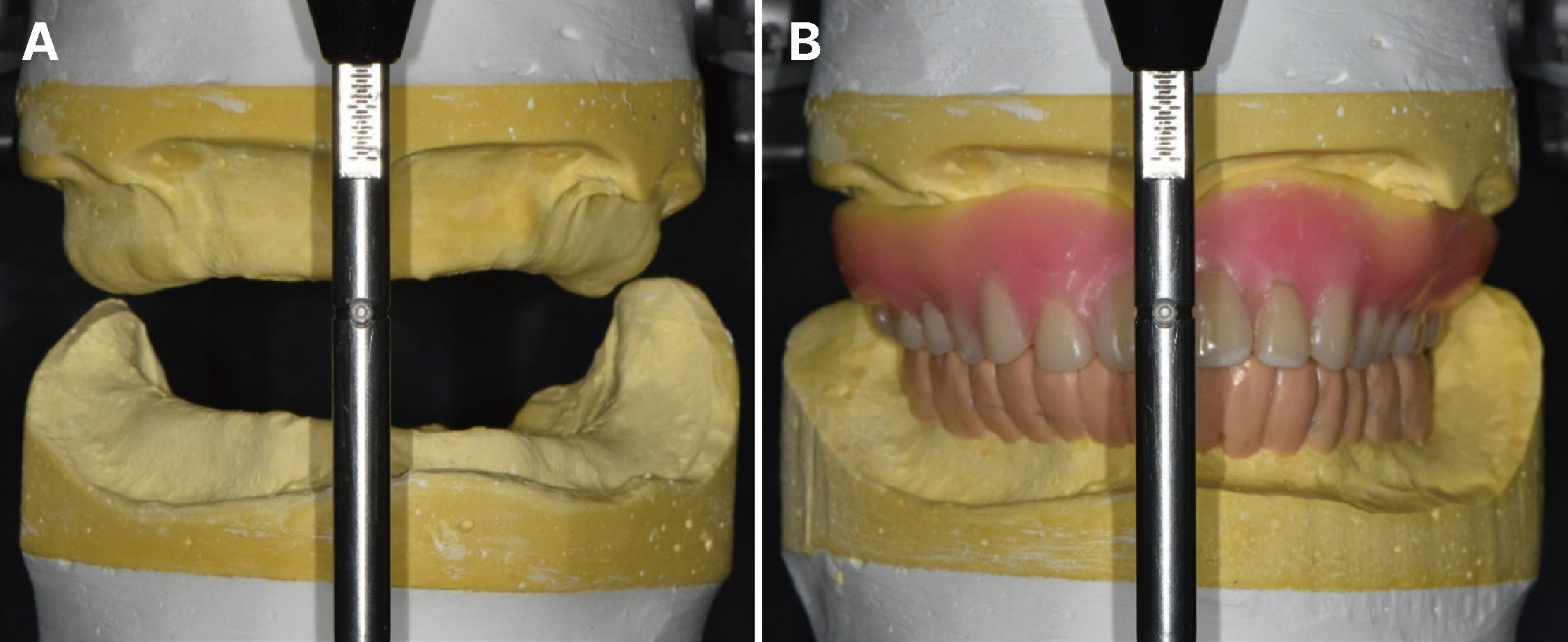
Fig. 3 Evaluation of diagnostic cast. (A) Mounting study cast on articulator, (B) Maxillary wax denture & Mandibular wax-up for fixed prosthesis.
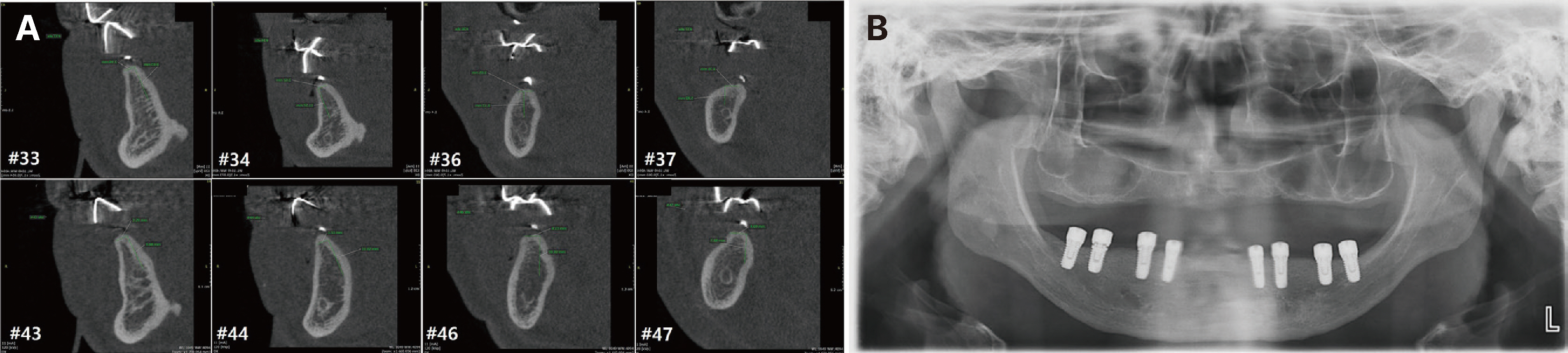
Fig. 4 Evaluation of radiographs for implant treatment. (A) Diagnosis of residual bone by CBCT, (B) Panoramic radiograph after implant placement.
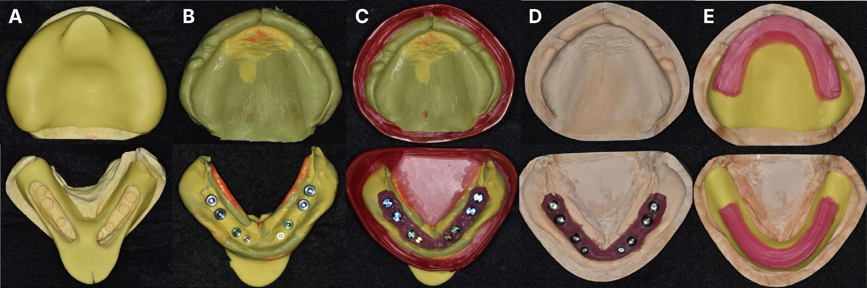
Fig. 5 (A) Fabrication of individual tray on study cast, (B) Final impression taking, (C) Beading and boxing, (D) Fabrication of master cast, (E) Fabrication of record base and occlusal rim.
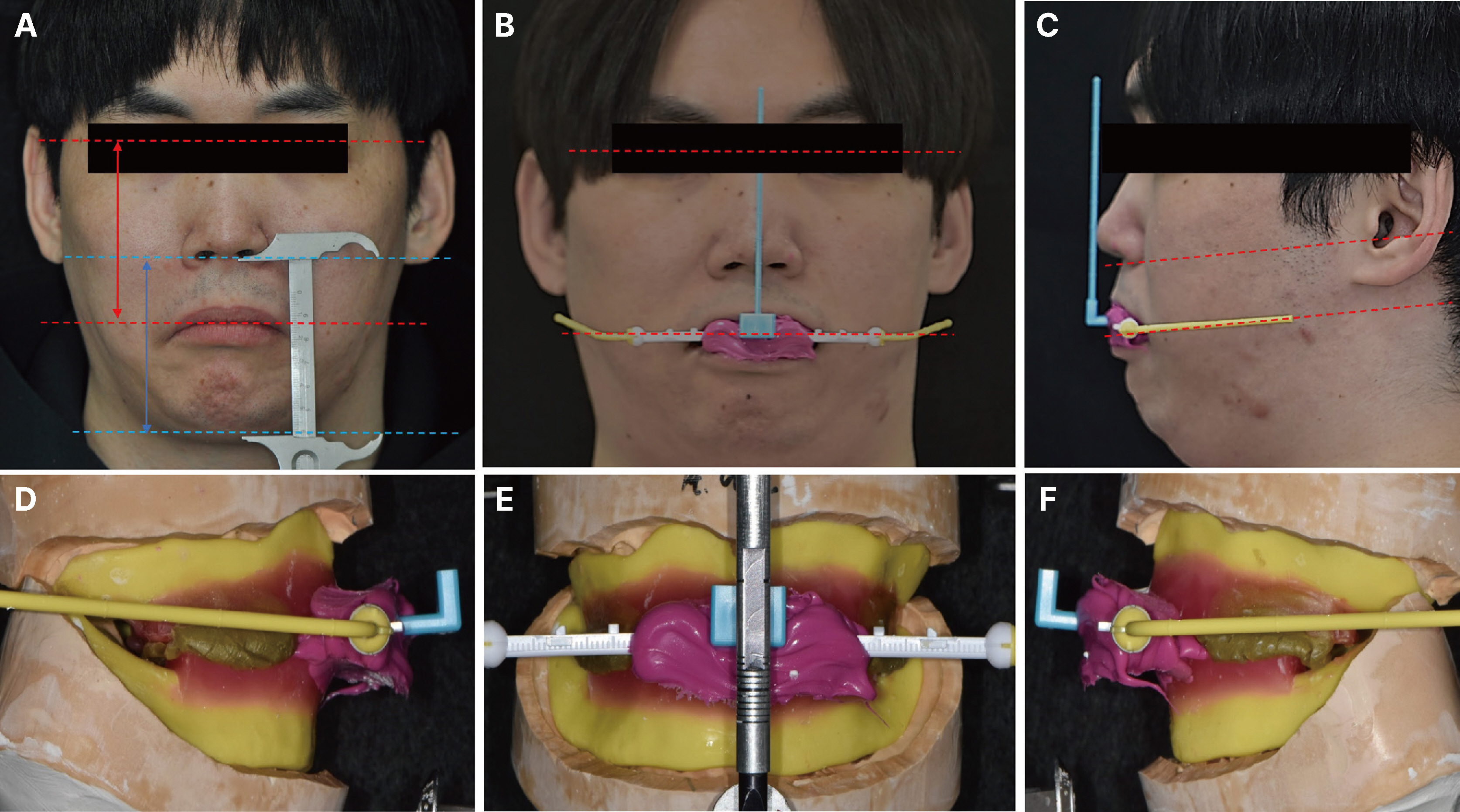
Fig. 6 Interocclusal relationship recording and transfer to articulator. (A) Determination of occlusal and vertical dimension, (B, C) Transfer of occlusal plane by POP bow system with interocclusal recording, (D - F) Mounting of maxillary and Mandibular master cast with occlusal rim attached POP bow system on articulator.
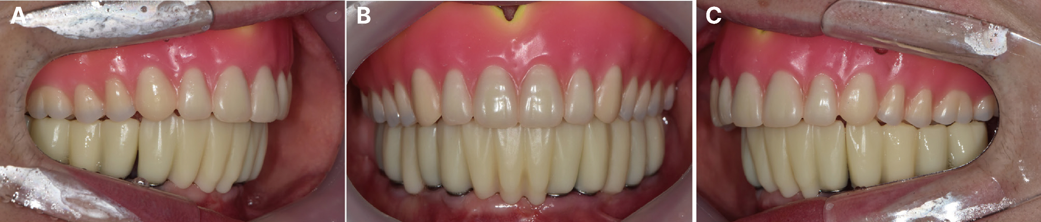
Fig. 7 Try-in of maxillary wax denture and mandibular provisional bridge with customized abutment. (A) Right lateral view, (B) Frontal view, (C) Left lateral view.
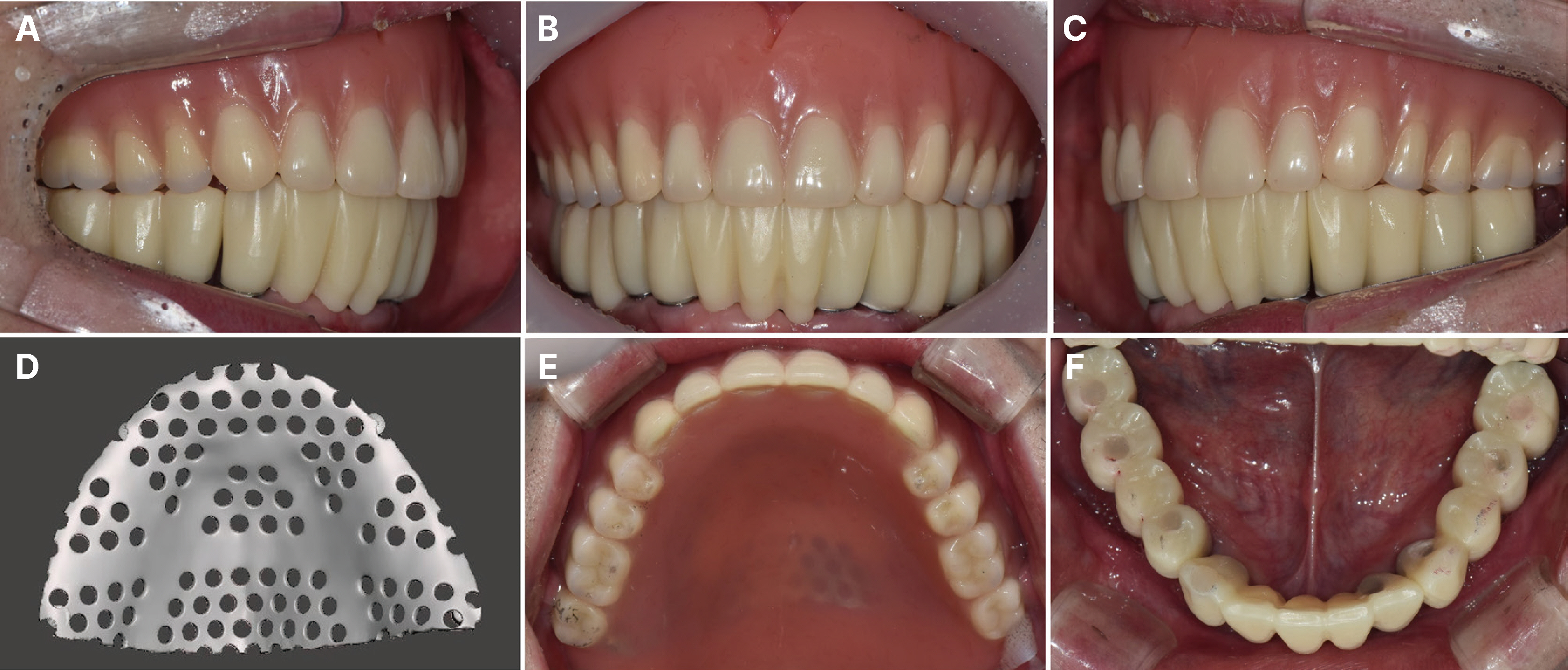
Fig. 8 Delivery of maxillary complete denture and mandibular implant provisional prosthesis. (A) Right lateral view, (B) Frontal view, (C) Left lateral view, (D) Design of 3D printing mesh framework at maxilla, (E) Occlusal view of maxilla, (F) Occlusal view of mandible.
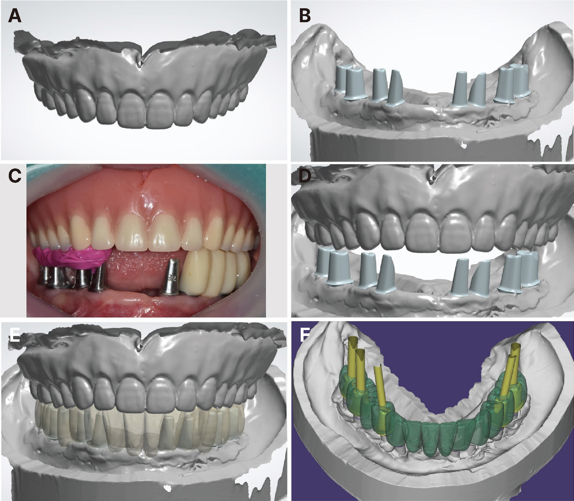
Fig. 9 Design of mandibular implant prosthesis. (A) Scan data of maxillary complete denture, (B) Scan data of mandibular customized titanium abutments, (C) After removing the temporary prosthesis on the right side of mandible, interocclusal recording was performed using a silicone bite table, followed by unilateral bite scanning, (D) Occlusal relation of maxilla and mandible was completed digitally by repeating the process while sequentially removing the three temporary prostheses, (E) Digital arrangement of mandibular fixed prosthesis, (F) Confirmation of implant screw hole path on mandibular fixed prosthesis.
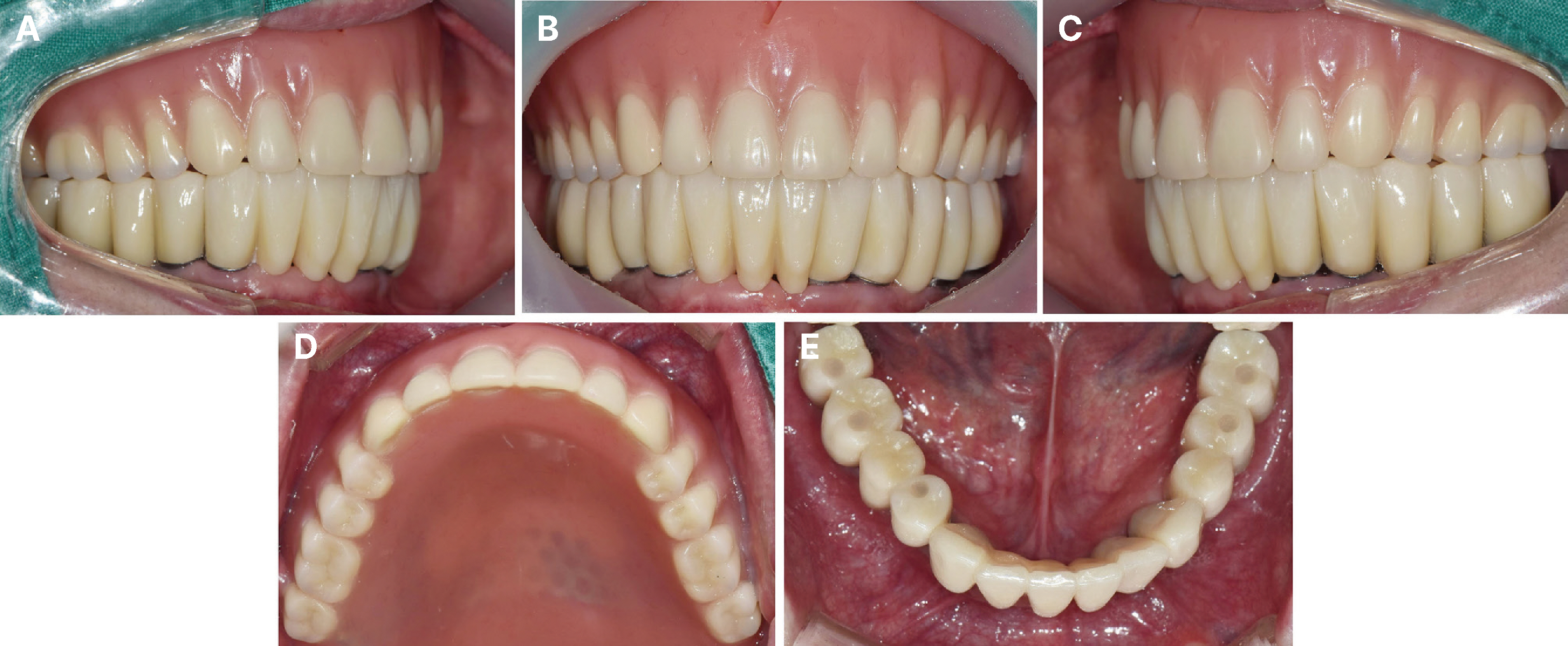
Fig. 10 Delivery of definite prosthesis on mandibular implant fixed prosthesis. (A) Right lateral view, (B) Frontal view, (C) Left lateral view, (D) Occlusal view of maxilla, (E) Occlusal view of mandible.
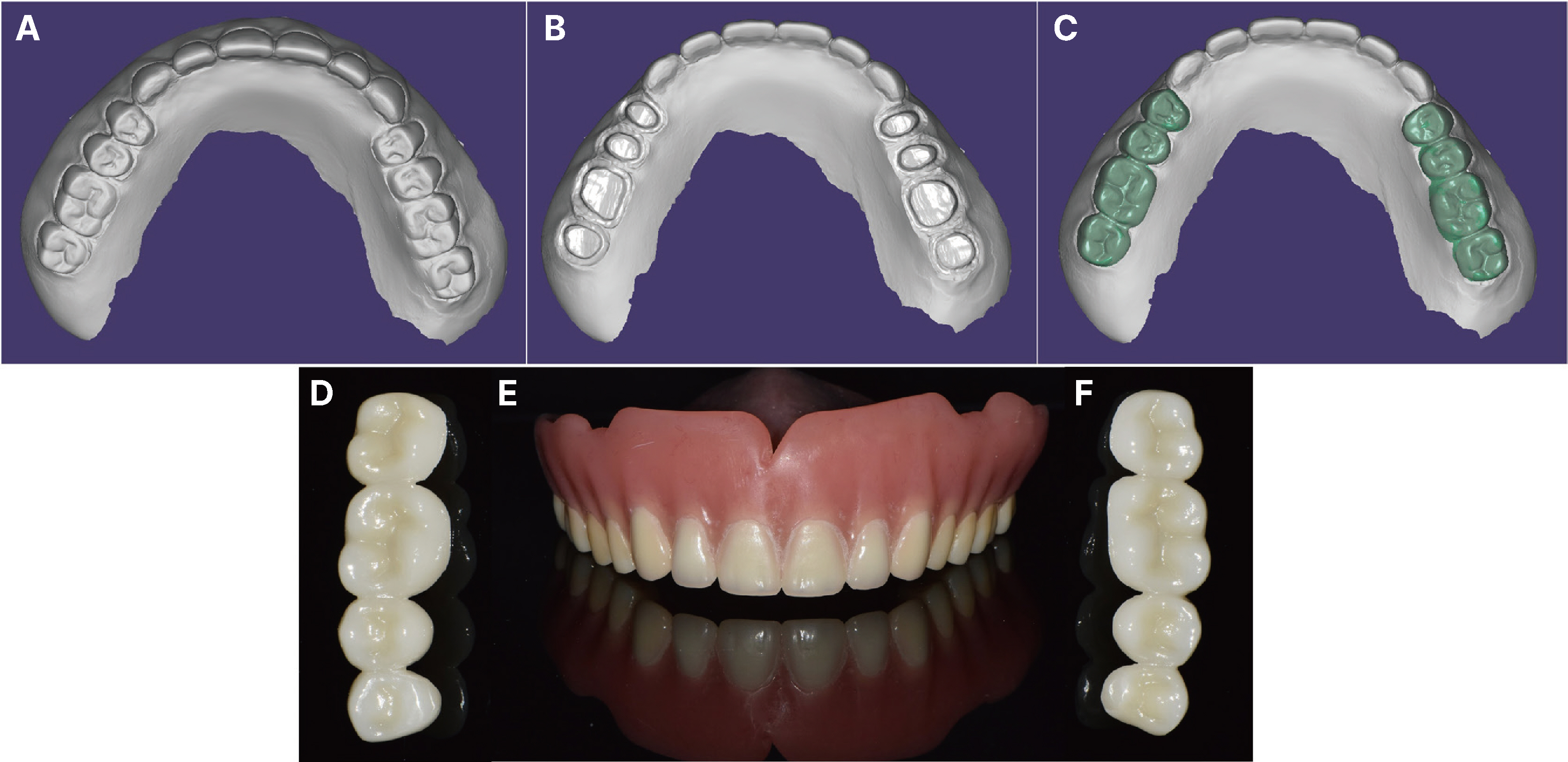
Fig. 11 (A) Scanning of maxillary complete denture, (B) Scanning after preparation of posterior artifcial teeth, (C) Design of a veneer-type zirconia occlusal surface by overlapping the (A) file with the (B) file, (D) Right posterior zirconia occlusion, (E) Maxillary complete denture with zirconia occlusion, (F) Left posterior zirconia occlusion.
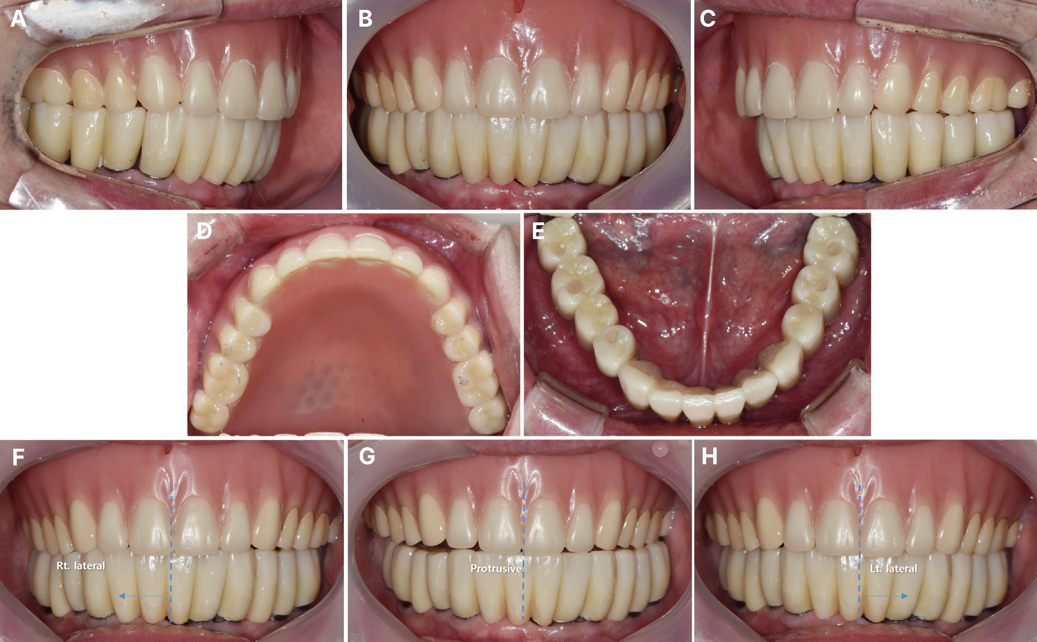
Fig. 12 After 6 months, maxillary complete denture with zirconia occlusion and mandibular implant fixed prosthesis. (A) Right lateral view, (B) Frontal view, (C) Left lateral view, (D) Maxillary occlusal view, (E) Mandibular occlusal view, (F) Right lateral movement, (G) Protrusive movement, (H) Left lateral movement.
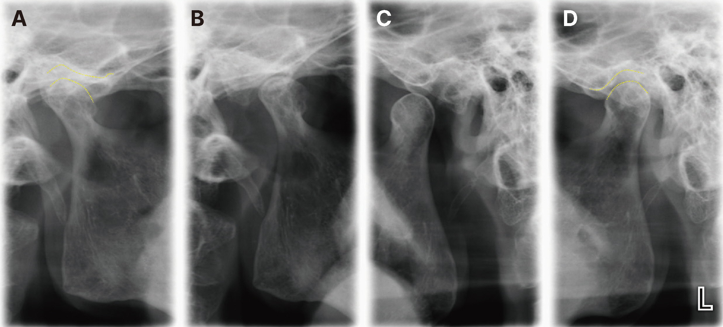
Fig. 13 Evaluation of TMJ (A) Right centric occlusion, (B) Right maximum opening, (C) Left maximum opening, (D) Left centric occlusion.
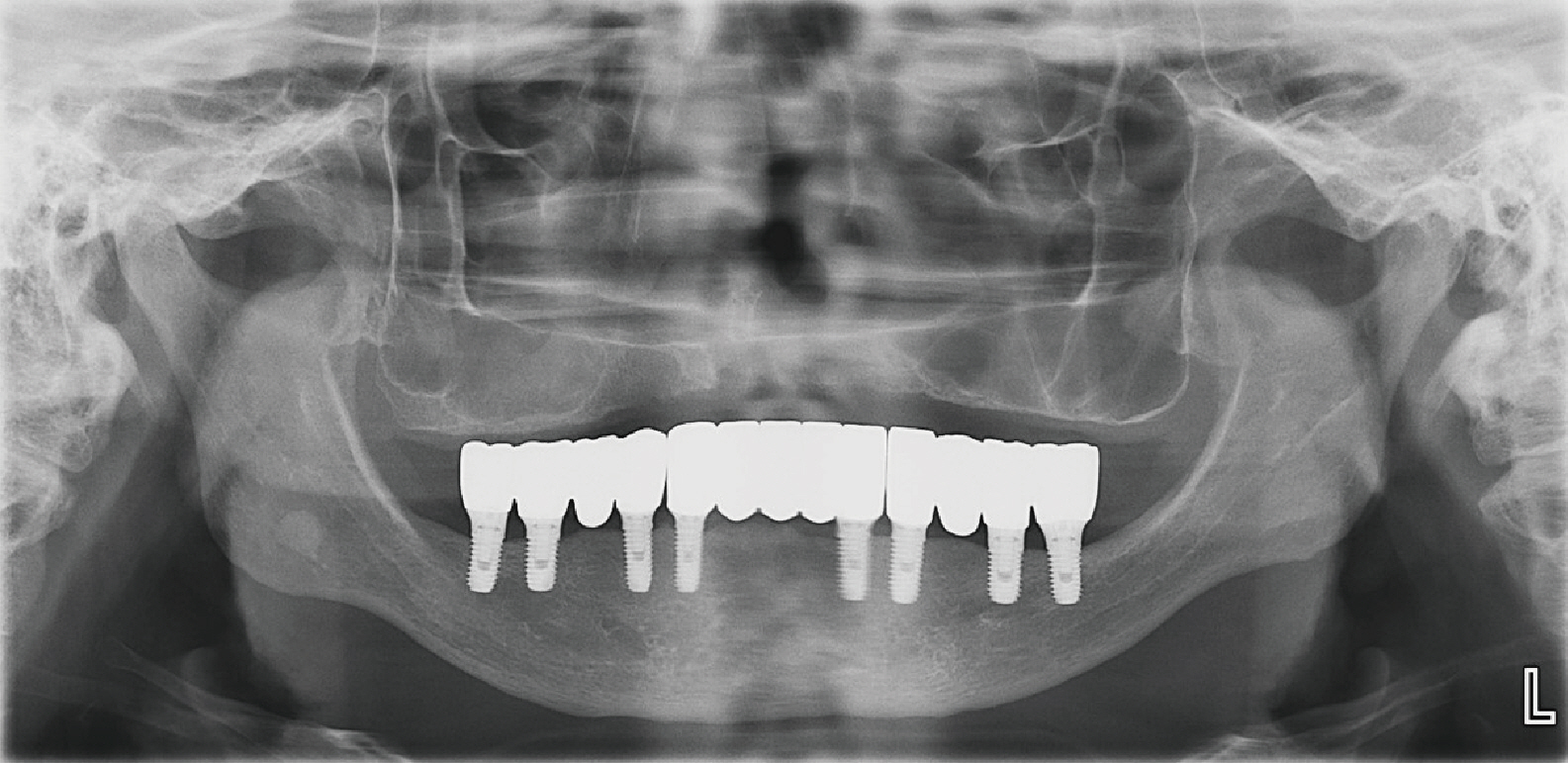
Fig. 14 Post-treatment panoramic radiograph.
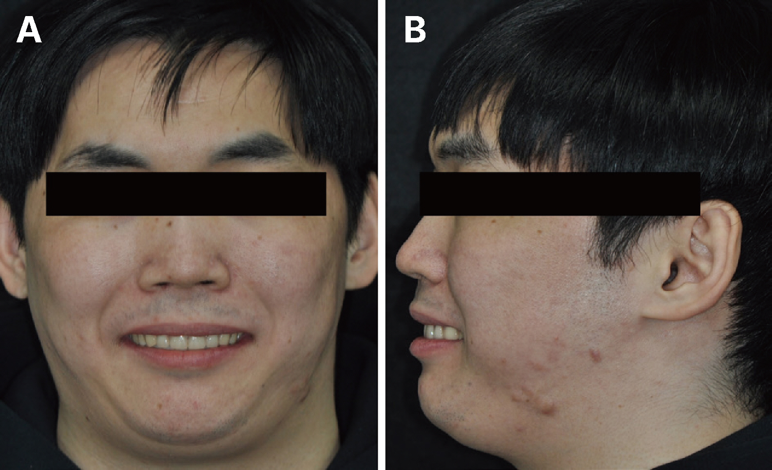
Fig. 15 Post-treatment extraoral view (A) Frontal smile view, (B) Lateral smile view.
Reference
-
References
1. Kai HL. 2007; Oromandibular dystonia. Oral Surg Oral Med Oral Pathol Oral Radio Endod. 104:491–6. DOI: 10.1016/j.tripleo.2007.04.001. PMID: 17689275.2. Blanchet PJ, Rompré PH, Lavigne GJ, Lamarche C. 2005; Oral dyskinesia: a clinical overview. Int J Prosthodont. 18:10–9. PMID: 15754887.3. Lumetti S, Ghiacci G, Macaluso GM, Amore M, Galli C, Calciolari E, Manfredi E. 2016; Tardive dyskinesia, oral parafunction, and implant-supported rehabilitation. Case Rep Dent. 2016:7167452. DOI: 10.1155/2016/7167452. PMID: 28050290. PMCID: PMC5168480. PMID: 83c8d830e50846ae801552e8235e6de1.4. Singh RK, Kundra S, Rani S. 2017; A prevalence based evaluation of etiology and site of fracture of acrylic resin dentures: a survey based original study. J Adv Med Dent Sci Res. 5:11–4.5. Wallace DH. 1964; The use of gold occlusal surfaces in complete and partial dentures. J Prosthet Dent. 14:326–33. DOI: 10.1016/0022-3913(64)90093-9.6. Balasubramaniam R, Ram S. 2008; Orofacial movement disorders. Oral Maxillofac Surg Clin North Am. 20:273–85. DOI: 10.1016/j.coms.2007.12.010. PMID: 18343330.7. Chee W, Jivraj S. 2006; Treatment planning of the edentulous mandible. Br Dent J. 201:337–47. DOI: 10.1038/sj.bdj.4814041. PMID: 16990883.8. Radke UM, Gundawar SM, Banarjee RS, Paldiwal AS. 2012; The single complete denture - a case report. Int J Clin Dent Sci. 3:76–81.9. Prachi G, Jyotsna S, Maya D, Milind K. 2018; Metal reinforced single complete denture - a case report. Int J Med Oral Res. 3:38–40.10. Diaz-Arnold AM, Vargas MA, Shaull KL, Laffoon JE, Qian F. 2008; Flexural and fatigue strengths of denture base resin. J Prosthet Dent. 100:47–51. DOI: 10.1016/S0022-3913(08)60136-5. PMID: 18589074.11. Upadhyay SR, Singh SV, Bhalla G, Kumar L, Singh BP. 2012; Modified functionally generated path technique for single complete denture against non-modified natural dentition. J Oral Biol Craniofac Res. 2:67–71. DOI: 10.1016/S2212-4268(12)60016-5. PMID: 25756037. PMCID: PMC3941280.12. Hu F, Pei Z, Wen Y. 2019; Using intraoral scanning technology for three-dimensional printing of Kennedy Class I removable partial denture metal framework: a clinical report. J Prosthodont. 28:e473–6. DOI: 10.1111/jopr.12712.13. McGuire MK, Nunn ME. 1996; Prognosis versus actual outcome. II. The effectiveness of clinical parameters in developing an accurate prognosis. J Periodontol. 67:658–65. DOI: 10.1902/jop.1996.67.7.658. PMID: 8832476.14. Örtorp A, Jönsson D, Mouhsen A, Vult von Steyern P. 2011; The fit of cobalt-chromium three-unit fixed dental prostheses fabricated with 4 different techniques: a comparative in vitro study. Dent Mater J. 27:356–63. DOI: 10.1016/j.dental.2010.11.015. PMID: 21163516.15. Cabianca M. 2003; Combination sydrome: treatment with dental implants. Implant Dent. 12:300–5. DOI: 10.1097/01.ID.0000094033.62974.BB. PMID: 14752966.16. Chong BJ, Thangavel AK, Rolton SB, Guazzato M, Klineberg IJ. 2015; Clinical and laboratory surface finishing procedures for zirconia on opposing human enamel wear: a laboratory study. J Mech Behav Biomed Mater. 50:93–103. DOI: 10.1016/j.jmbbm.2015.06.007. PMID: 26116957.17. Ghazal M, Kern M. 2009; The influence of antagonistic surface roughness on the wear of human enamel and nanofilled composite resin artificial teeth. J Prosthet Dent. 101:342–9. DOI: 10.1016/S0022-3913(09)60068-8. PMID: 19410068.18. Ghazal M, Yang B, Ludwig K, Kern M. 2008; Two-body wear of resin and ceramic denture teeth in comparison to human enamel. Dent Mater. 24:502–7. DOI: 10.1016/j.dental.2007.04.012. PMID: 17688934.19. Shori K, Shori T, Shori D, Chavan R. 2015; Achieving esthetic perfection by zirconia: A Case Report. Int J Dent Med Res. 1:146–9.20. Lawson NC, Janyavula S, Syklawer S, Mclaren EA, Burgess JO. 2014; Wear of enamel opposing zirconia and lithium disilicate after adjustment, polishing and glazing. J Dent. 42:1586–91. DOI: 10.1016/j.jdent.2014.09.008. PMID: 25257823.21. Lambert H, Durand JC, Jacquot B, Fages M. 2017; Dental biomaterials for chairside CAD-CAM: State of the art. J Adv Prosthodont. 9:486–95. DOI: 10.4047/jap.2017.9.6.486. PMID: 29279770. PMCID: PMC5741454.22. Gibson RM. 1989; Smiling and Facial Exercise. Dent Clin North Am. 33:139–44. DOI: 10.1016/S0011-8532(22)01182-X. PMID: 2721794.
- Full Text Links
-
- Actions
-
Cited
- CITED
-
- Close
- Share
-
- Similar articles
-
- Comparison of treatments for maxillary full denture and mandibular implant-supported fixed prosthesis in completely edentulous patients: A case report
- Implant Surgery for Fixed Implant-supported Prostheses in the Edentulous Mandible: A Case Report
- Rehabilitation of maxillary partial edentulous patients using implant assisted removable partial denture
- Treatment with upper complete denture and lower implant-fixed restorations on an elderly patient presenting fully edentulous maxilla and bilateral posterior edentulous mandible: a case report
- Restoration of bilateral distal extension removable partial denture using a fixed implant prosthesis in unilateral partial edentulous patient: A case report
