Restor Dent Endod.
2022 Nov;47(4):e39. 10.5395/rde.2022.47.e39.
The prevalence and characteristics of external cervical resorption based on cone-beam computed tomographic imaging: a cross-sectional study
- Affiliations
-
- 1Division of Oral Radiology, Department of Oral Diagnosis, Piracicaba Dental School, University of Campinas (UNICAMP), Sao Paulo, Brazil
- 2Department of Oral Diagnosis, São Paulo Dental School, University of São Paulo (USP), São Paulo, Brazil
- KMID: 2548143
- DOI: http://doi.org/10.5395/rde.2022.47.e39
Abstract
Objectives
This study investigated the prevalence and characteristics of external cervical resorption (ECR) regarding sex, age, tooth, stages of progression, and portal of entry, using cone-beam computed tomography (CBCT) scans.
Materials and Methods
CBCT scans of 1,313 patients from a Brazilian subpopulation comprising 883 female and 430 male patients (mean age, 55.2 years), acquired using a PreXion 3D CBCT unit, were evaluated. All permanent teeth included in the scans were evaluated for the presence of ECR according to the 3-dimensional classification and the portal of entry. The association between the presence of ECR and the factors studied was assessed using the χ2 test. Intra-observer agreement was analyzed with the kappa test (α = 0.05).
Results
In total, 6,240 teeth were analyzed, of which 84 (1.35%) were affected by ECR. A significant association was found between the presence of ECR and sex, with a higher prevalence in male patients (p = 0.002). The most frequently affected teeth were the mandibular and maxillary central incisors. The most common height was the mid-third of the root. For the portal of entry, 44% of cases were on the proximal surfaces, 40.5% on the lingual/palatal surface and 15.5% on the buccal surface. Intra-observer agreement was excellent.
Conclusions
The prevalence of ECR was 1.35%, with a higher prevalence in male patients and a wide age distribution. The mandibular and maxillary central incisors were the most commonly affected teeth, and cases of ECR most frequently showed a height into the midthird of the root and proximal entry.
Figure
-
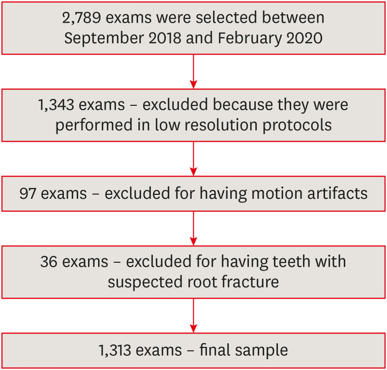
Figure 1 Flowchart showing the sample selection process.
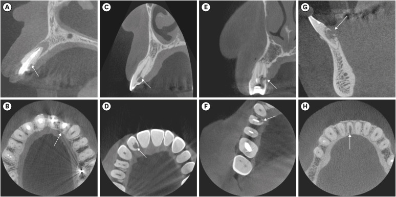
Figure 2 Examples of external cervical resorption indicated by white arrows. (A, B) Classification 1Ad. (C, D) classification 2Bd. (E, F) classification 3Ap. (G, H) classification 4Ap, according to the 3-dimensional classification.
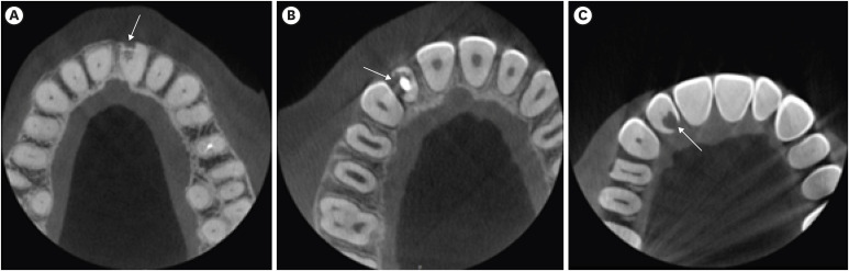
Figure 3 Examples of the portal of entry of external cervical resorption lesions indicated by white arrows. (A) buccal, (B) proximal, (C) palatal.
Reference
-
1. Kandalgaonkar SD, Gharat LA, Tupsakhare SD, Gabhane MH. Invasive cervical resorption: a review. J Int Oral Health. 2013; 5:124–130.2. Patel S, Mavridou AM, Lambrechts P, Saberi N. External cervical resorption-part 1: histopathology, distribution and presentation. Int Endod J. 2018; 51:1205–1223. PMID: 29704466.
Article3. Vaz de Souza D, Schirru E, Mannocci F, Foschi F, Patel S. External cervical resorption: a comparison of the diagnostic efficacy using 2 different cone-beam computed tomographic units and periapical radiographs. J Endod. 2017; 43:121–125. PMID: 27939734.
Article4. Irinakis E, Aleksejuniene J, Shen Y, Haapasalo M. External cervical resorption: a retrospective case-control study. J Endod. 2020; 46:1420–1427. PMID: 32525057.5. Matny LE, Ruparel NB, Levin MD, Noujeim M, Diogenes A. A volumetric assessment of external cervical resorption cases and its correlation to classification, treatment planning, and expected prognosis. J Endod. 2020; 46:1052–1058. PMID: 32437787.
Article6. Heithersay GS. Invasive cervical resorption. Endod Topics. 2004; 7:73–92.
Article7. Chen Y, Huang Y, Deng X. A review of external cervical resorption. J Endod. 2021; 47:883–894. PMID: 33745945.
Article8. Goodell KB, Mines P, Kersten DD. Impact of cone-beam computed tomography on treatment planning for external cervical resorption and a novel axial slice-based classification system. J Endod. 2018; 44:239–244. PMID: 29229454.
Article9. Mavridou AM, Bergmans L, Barendregt D, Lambrechts P. Descriptive analysis of factors associated with external cervical resorption. J Endod. 2017; 43:1602–1610. PMID: 28807370.
Article10. Heithersay GS. Invasive cervical resorption: an analysis of potential predisposing factors. Quintessence Int. 1999; 30:83–95. PMID: 10356560.11. Suhr Villefrance J, Kirkevang LL, Wenzel A, Væth M, Matzen LH. Impact of cone beam CT on diagnosis of external cervical resorption: the severity of resorption assessed in periapical radiographs and cone beam CT. A prospective clinical study. Dentomaxillofac Radiol. 2022; 51:20210279. PMID: 34520244.
Article12. Patel S, Foschi F, Mannocci F, Patel K. External cervical resorption: a three-dimensional classification. Int Endod J. 2018; 51:206–214. PMID: 28746776.
Article13. Rotondi O, Waldon P, Kim SG. The disease process, diagnosis and treatment of invasive cervical resorption: a review. Dent J. 2020; 8:64.
Article14. Jebril A, Aljamani S, Jarad F. The surgical management of external cervical resorption: a retrospective observational study of treatment outcomes and classifications. J Endod. 2020; 46:778–785. PMID: 32334857.
Article15. Jeng PY, Lin LD, Chang SH, Lee YL, Wang CY, Jeng JH, Tsai YL. Invasive cervical resorption—distribution, potential predisposing factors, and clinical characteristics. J Endod. 2020; 46:475–482. PMID: 32115249.
Article16. Landis JR, Koch GG. The measurement of observer agreement for categorical data. Biometrics. 1977; 33:159–174. PMID: 843571.
Article17. Patel J, Beddis HP. How to assess and manage external cervical resorption. Br Dent J. 2019; 227:695–701. PMID: 31654002.
Article18. Thönen A, Peltomäki T, Patcas R, Zehnder M. Occurrence of cervical invasive root resorption in first and second molar teeth of orthodontic patients eight years after bracket removal. J Endod. 2013; 39:27–30. PMID: 23228253.
Article19. AAE and AAOMR joint position statement: use of cone beam computed tomography in endodontics 2015 update. J Endod. 2015; 41:1393–1396. PMID: 26320105.20. Patel K, Mannocci F, Patel S. The assessment and management of external cervical resorption with periapical radiographs and cone-beam computed tomography: a clinical study. J Endod. 2016; 42:1435–1440. PMID: 27507628.
Article
- Full Text Links
-
- Actions
-
Cited
- CITED
-
- Close
- Share
-
- Similar articles
-
- Multiple idiopathic external and internal resorption: Case report with cone-beam computed tomography findings
- Three-dimensional imaging modalities in endodontics
- Cone-beam computed tomographic reconstructions in the evaluation of maxillary impacted canines
- CBCT findings of periapical cemento-osseous dysplasia: A case report
- Cone beam CT findings of retromolar canals: Report of cases and literature review
