Restor Dent Endod.
2021 Nov;46(4):e57. 10.5395/rde.2021.46.e57.
Porosity and pore size distribution in high-viscosity and conventional glass ionomer cements: a micro-computed tomography study
- Affiliations
-
- 1Department of Pediatric Dentistry and Orthodontics, Universidade Federal do Rio de Janeiro – UFRJ, School of Dentistry, Rio de Janeiro, RJ, Brazil
- 2Laboratory of Nuclear Instrumentation, Universidade Federal do Rio de Janeiro – UFRJ, Rio de Janeiro, RJ, Brazil
- KMID: 2548101
- DOI: http://doi.org/10.5395/rde.2021.46.e57
Abstract
Objectives
This study aimed to compare and evaluate the porosity and pore size distribution of high-viscosity glass ionomer cements (HVGICs) and conventional glass ionomer cements (GICs) using micro-computed tomography (micro-CT).
Materials and Methods
Forty cylindrical specimens (n = 10) were produced in standardized molds using HVGICs and conventional GICs (Ketac Molar Easymix, Vitro Molar, MaxxionR, and Riva Self-Cure). The specimens were prepared according to ISO 9917-1 standards, scanned in a high-energy micro-CT device, and reconstructed using specific parameters. After reconstruction, segmentation procedures, and image analysis, total porosity and pore size distribution were obtained for specimens in each group. After checking the normality of the data distribution, the Kruskal-Wallis test followed by the Student-Newman-Keuls test was used to detect differences in porosity among the experimental groups with a 5% significance level.
Results
Ketac Molar Easymix showed statistically significantly lower total porosity (0.15%) than MaxxionR (0.62%), Riva (0.42%), and Vitro Molar (0.57%). The pore size in all experimental cements was within the small-size range (< 0.01 mm3 ), but Vitro Molar showed statistically significantly more pores/defects with a larger size (> 0.01 mm3 ).
Conclusions
Major differences in porosity and pore size were identified among the evaluated GICs. Among these, the Ketac Molar Easymix HVGIC showed the lowest porosity and void size.
Figure
-
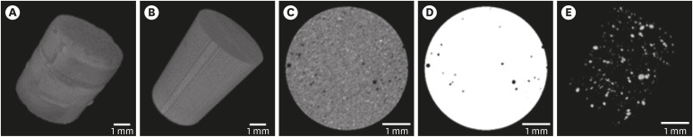
Figure 1 Glass ionomer cement specimens and porosity. (A) Original specimen. (B) Specimen after definition of the volume of interest. (C) Cross-sectional slice of the specimen after normalization and filtering. (D) Application of the iterative threshold. (E) Three-dimensional renderization of pore segmentation.
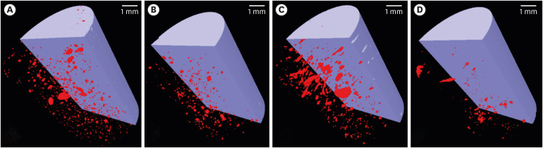
Figure 2 Three-dimensional volume rendering obtained after image acquisition and reconstruction of a representative specimen of each studied material. (A) MaxxionR; (B) Riva Self-Cure; (C) Vitro Molar; (D) Ketac Molar Easymix.
Reference
-
1. Sidhu SK, Nicholson JW. A review of glass-ionomer cements for clinical dentistry. J Funct Biomater. 2016; 7:16.
Article2. Coutinho E, Cardoso MV, De Munck J, Neves AA, Van Landuyt KL, Poitevin A, Peumans M, Lambrechts P, Van Meerbeek B. Bonding effectiveness and interfacial characterization of a nano-filled resin-modified glass-ionomer. Dent Mater. 2009; 25:1347–1357. PMID: 19595446.
Article3. Berg JH, Croll TP. Glass ionomer restorative cement systems: an update. Pediatr Dent. 2015; 37:116–124. PMID: 25905652.4. Hesse D, Bonifácio CC, Bönecker M, Guglielmi Cde A, da Franca C, van Amerongen WE, Colares V, Raggio DP. Survival rate of Atraumatic Restorative Treatment (ART) restorations using a glass ionomer bilayer technique with a nanofilled coating: a Bi-center randomized clinical trial. Pediatr Dent. 2016; 38:18–24.5. Burke FJT, Lucarotti PSK. The ultimate guide to restoration longevity in England and Wales. Part 3: glass ionomer restorations - time to next intervention and to extraction of the restored tooth. Br Dent J. 2018; 224:865–874. PMID: 29855590.
Article6. Frankenberger R, Sindel J, Krämer N. Viscous glass-ionomer cements: a new alternative to amalgam in the primary dentition? Quintessence Int. 1997; 28:667–676. PMID: 9477887.7. Schwendicke F, Göstemeyer G, Blunck U, Paris S, Hsu LY, Tu YK. Directly placed restorative materials: Review and network meta-analysis. J Dent Res. 2016; 95:613–622. PMID: 26912220.8. van Dijken JW, Pallesen U. Fracture frequency and longevity of fractured resin composite, polyacid-modified resin composite, and resin-modified glass ionomer cement class IV restorations: an up to 14 years of follow-up. Clin Oral Investig. 2010; 14:217–222.
Article9. Leal S, Bonifacio C, Raggio D, Frencken J. Atraumatic restorative treatment: restorative component. Monogr Oral Sci. 2018; 27:92–102. PMID: 29794453.
Article10. Hilgert LA, de Amorim RG, Leal SC, Mulder J, Creugers NH, Frencken JE. Is high-viscosity glass-ionomer-cement a successor to amalgam for treating primary molars? Dent Mater. 2014; 30:1172–1178. PMID: 25132283.
Article11. de Amorim RG, Frencken JE, Raggio DP, Chen X, Hu X, Leal SC. Survival percentages of atraumatic restorative treatment (ART) restorations and sealants in posterior teeth: an updated systematic review and meta-analysis. Clin Oral Investig. 2018; 22:2703–2725.
Article12. Nomoto R, McCabe JF. Effect of mixing methods on the compressive strength of glass ionomer cements. J Dent. 2001; 29:205–210. PMID: 11306162.
Article13. Nimmo JR. Porosity and pore size distribution. Reference module in earth systems and environmental sciences. Amsterdam: Elsevier;2013.14. Mitchell CA, Douglas WH. Comparison of the porosity of hand-mixed and capsulated glass-ionomer luting cements. Biomaterials. 1997; 18:1127–1131. PMID: 9247351.
Article15. Nicholson JW. Maturation processes in glass-ionomer dental cements. Acta Biomater Odontol Scand. 2018; 4:63–71. PMID: 30083577.
Article16. Geirsson J, Thompson JY, Bayne SC. Porosity evaluation and pore size distribution of a novel directly placed ceramic restorative material. Dent Mater. 2004; 20:987–995. PMID: 15501328.
Article17. Coldebella CR, Santos-Pinto L, Zuanon AC. Effect of ultrasonic excitation on the porosity of glass ionomer cement: a scanning electron microscope evaluation. Microsc Res Tech. 2011; 74:54–57. PMID: 21181710.
Article18. Covey DA, Ewoldsen NO. Porosity in manually and machine mixed resin-modified glass ionomer cements. Oper Dent. 2001; 26:617–623. PMID: 11699187.19. Malkoç MA, Sevimay M, Tatar İ, Çelik HH. Micro-CT detection and characterization of porosity in luting cements. J Prosthodont. 2015; 24:553–561. PMID: 25557068.
Article20. Nomoto R, Komoriyama M, McCabe JF, Hirano S. Effect of mixing method on the porosity of encapsulated glass ionomer cement. Dent Mater. 2004; 20:972–978. PMID: 15501326.
Article21. De Souza ET, Nunes Tameirão MD, Roter JM, De Assis JT, De Almeida Neves A, De-Deus GA. Tridimensional quantitative porosity characterization of three set calcium silicate-based repair cements for endodontic use. Microsc Res Tech. 2013; 76:1093–1098. PMID: 23913667.
Article22. Faul F, Erdfelder E, Lang AG, Buchner A. G*Power 3: a flexible statistical power analysis program for the social, behavioral, and biomedical sciences. Behav Res Methods. 2007; 39:175–191. PMID: 17695343.
Article23. Calvo AF, Kicuti A, Tedesco TK, Braga MM, Raggio DP. Evaluation of the relationship between the cost and properties of glass ionomer cements indicated for atraumatic restorative treatment. Braz Oral Res. 2016; 30:S1806-83242016000100201.
Article24. Schmid B, Schindelin J, Cardona A, Longair M, Heisenberg M. A high-level 3D visualization API for Java and ImageJ. BMC Bioinformatics. 2010; 11:274. PMID: 20492697.
Article25. Anna Luisa de Brito P, Isabel Cristina O, Clarissa Calil B, Ana Flávia Bissoto C, José Carlos Pettorossi I, Daniela Prócida R. One year survival rate of Ketac Molar versus Vitro Molar for occlusoproximal ART restorations: a RCT. Braz Oral Res. 2017; 31:e88. PMID: 29116299.
Article26. Bonifácio CC, Hesse D, Raggio DP, Bönecker M, van Loveren C, van Amerongen WE. The effect of GIC-brand on the survival rate of proximal-ART restorations. Int J Paediatr Dent. 2013; 23:251–258. PMID: 22891625.
Article27. Benetti AR, Jacobsen J, Lehnhoff B, Momsen NC, Okhrimenko DV, Telling MT, Kardjilov N, Strobl M, Seydel T, Manke I, Bordallo HN. How mobile are protons in the structure of dental glass ionomer cements? Sci Rep. 2015; 5:8972. PMID: 25754555.
Article28. Peez R, Frank S. The physical-mechanical performance of the new Ketac Molar Easymix compared to commercially available glass ionomer restoratives. J Dent. 2006; 34:582–587. PMID: 16581174.
Article29. Raggio DP, Bonifácio CC, Bönecker M, Imparato JC, Gee AJ, Amerongen WE. Effect of insertion method on Knoop hardness of high viscous glass ionomer cements. Braz Dent J. 2010; 21:439–445. PMID: 21180801.
Article30. Neves AB, Bergstrom TG, Fonseca-Gonçalves A, Dos Santos TMP, Lopes RT, de Almeida Neves A. Mineral density changes in bovine carious dentin after treatment with bioactive dental cements: a comparative micro-CT study. Clin Oral Investig. 2019; 23:1865–1870.
Article31. Bonifácio CC, Kleverlaan CJ, Raggio DP, Werner A, de Carvalho RC, van Amerongen WE. Physical-mechanical properties of glass ionomer cements indicated for atraumatic restorative treatment. Aust Dent J. 2009; 54:233–237. PMID: 19709111.
Article32. Nicholson J, Czarnecka B. Conventional glass-ionomer cements. Nicholson J, Czarnecka B, editors. Materials for the direct restoration of teeth. Amsterdam: Elsevier;2016. p. 107–136.33. Santa G, Bentz D, Weiss J. Capillary porosity depercolation in cement-based materials: Measurement techniques and factors which influence their interpretation. Cement Concr Res. 2011; 41:854–864.
Article
- Full Text Links
-
- Actions
-
Cited
- CITED
-
- Close
- Share
-
- Similar articles
-
- Comparison of capsule type resin modified glass ionomer porosity according to mixing methods
- In vitro study on the fluoride release from glass ionomer cements and a fluoride-containing resin
- Comparative studies on the retentive values of various dental cements used to retain orthodontic bands
- Current aspects and prospects of glass ionomer cements for clinical dentistry
- The effects of surface treatments on shear bond strengths of light-cured and chemically cured glass ionomer cements to enamel
