The double-barrier technique using platelet-rich fibrin for closure of oroantral fistulas
- Affiliations
-
- 1Department of Oral and Maxillofacial Surgery, Kyung Hee University Dental Hospital at Gangdong, Kyung Hee University College of Dentistry, Seoul, Korea
- KMID: 2544320
- DOI: http://doi.org/10.5125/jkaoms.2023.49.3.163
Abstract
- An oroantral fistula (OAF) or oroantral communication (OAC) is an opening between the oral cavity and the maxillary sinus. If left untreated, these openings may cause chronic maxillary sinusitis. Although small defects (diameter <5 mm) may close spontaneously, larger communications require surgical intervention. Various studies have been conducted on OAC closure using a platelet-rich fibrin (PRF) membrane; most of these prior studies have involved simple direct application of PRF clots. This study introduces a new “double-barrier technique” using PRF for closure of an OAF involving sinus mucosal lifting and closure. The PRF material is inserted into the prepared maxillary sinus space, and the buccal advancement flap covers the oral side. This technique was successfully used to treat two patients with chronic OAF in the posterior maxillary region after implant removal or tooth extraction. The use of a PRF membrane in a double-barrier technique may have advantages in soft-tissue healing and could enable easy closure of chronic OAF with minimal trauma.
Keyword
Figure
-
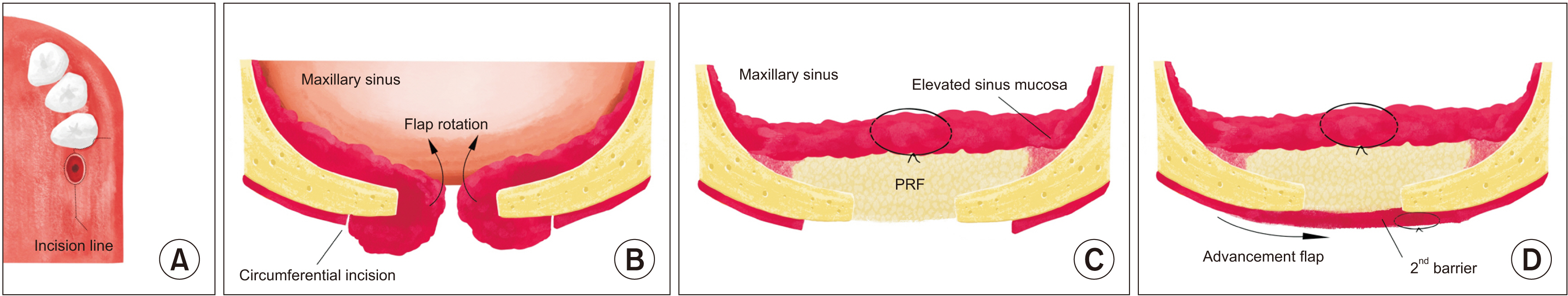
Fig. 1 A schematic of the incision line (A), antral flap rotation and elevation as the first barrier (B), platelet-rich fibrin (PRF) insertion into the prepared maxillary sinus space (C), a buccal advancement flap as the second barrier (D).
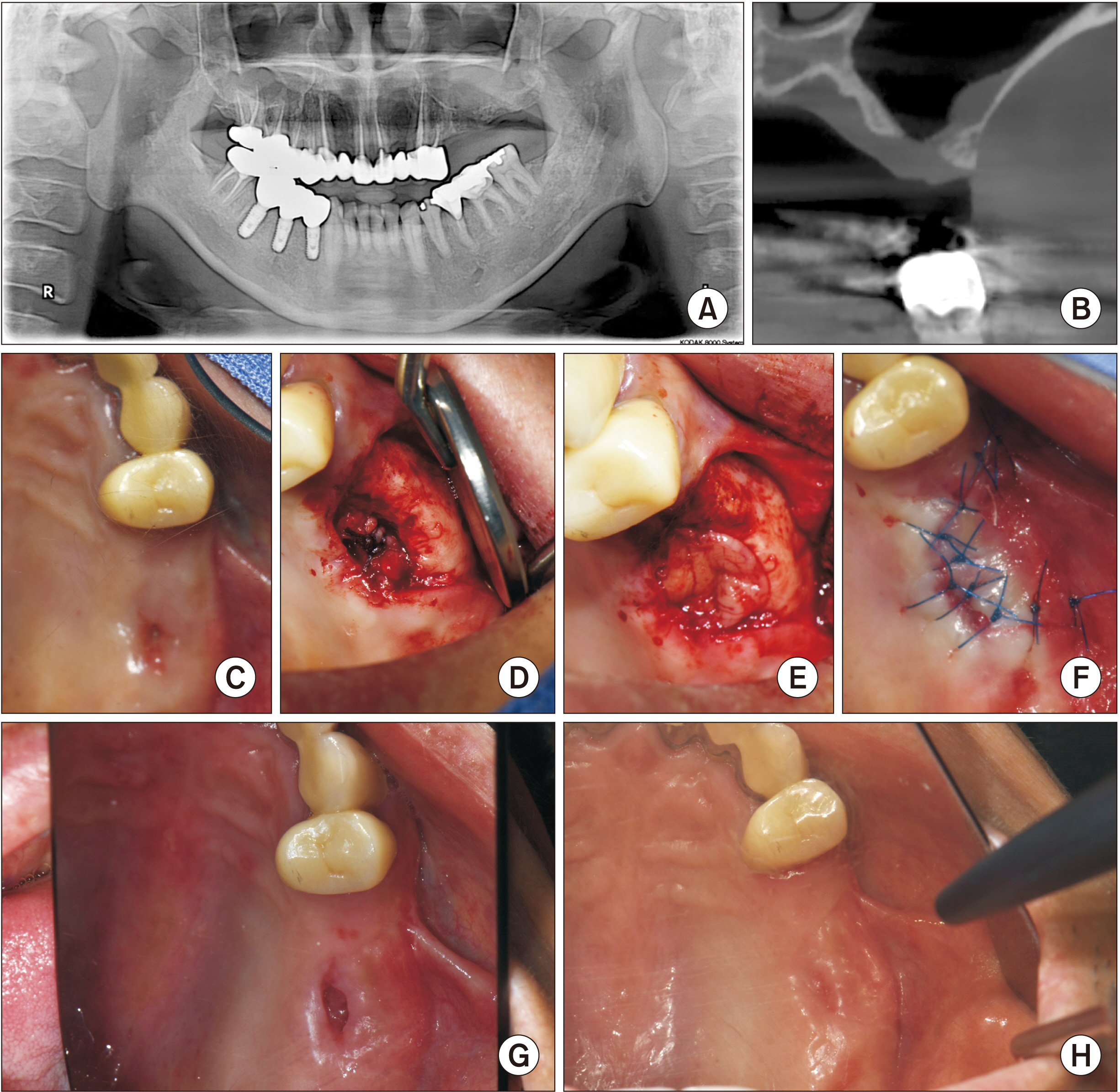
Fig. 2 A. A pre-surgery panoramic radiograph of Patient No. 1. B. The cone-beam computed tomography view. C. A view of the oroantral fistula from the oral side. D. The view of the elevated and sutured sinus mucosa. E. A view of the platelet-rich fibrin clots inserted as a membrane. F. The view of the sutured buccal advancement flap. G. A view from the oral side at two weeks postoperatively. H. A view from the oral side at two months after surgery.

Fig. 3 A. A pre-surgery panoramic radiographs of Patient No. 2. B. The cone-beam computed tomography view. C. A view of the oroantral fistula from the oral side. D. The view of the elevated sinus mucosa. E. A view of the platelet-rich fibrin clots inserted as a membrane. F. The view of the sutured buccal advancement flap. G. A view from the oral side at two weeks postoperatively. H. A view from the oral side at two months after surgery.
Reference
-
References
1. Kim MK, Han W, Kim SG. 2017; The use of the buccal fat pad flap for oral reconstruction. Maxillofac Plast Reconstr Surg. 39:5. https://doi.org/10.1186/s40902-017-0105-5. DOI: 10.1186/s40902-017-0105-5. PMID: 28286743. PMCID: PMC5325802.
Article2. Bilginaylar K. 2018; The use of platelet-rich fibrin for immediate closure of acute oroantral communications: an alternative approach. J Oral Maxillofac Surg. 76:278–86. https://doi.org/10.1016/j.joms.2017.07.168. DOI: 10.1016/j.joms.2017.07.168. PMID: 28859924.
Article3. Cheng GL, Tatakis DN. 2020; Collagen strip technique: a novel approach for ridge preservation and concomitant oroantral communication management after implant explantation. Clin Adv Periodontics. 10:135–9. https://doi.org/10.1002/cap.10092. DOI: 10.1002/cap.10092. PMID: 32065734.
Article4. Noel JE, Teo NW, Divi V, Nayak JV. 2016; Use of pedicled nasoseptal flap for pathologic oroantral fistula closure. J Oral Maxillofac Surg. 74:704.e1–6. https://doi.org/10.1016/j.joms.2015.11.010. DOI: 10.1016/j.joms.2015.11.010. PMID: 26704432.
Article5. Demetoglu U, Ocak H, Bilge S. 2018; Closure of oroantral communication with plasma-rich fibrin membrane. J Craniofac Surg. 29:e367–70. https://doi.org/10.1097/scs.0000000000004360. DOI: 10.1097/SCS.0000000000004360. PMID: 29485557.
Article6. Lee CYS. 2019; Closure of an oroantral communication using leucocyte-platelet rich fibrin: a novel technique using regenerative medicine. J Dent Maxillofac Surg. 2:101–6. https://doi.org/10.18314/jdms.v2i1.1540. DOI: 10.18314/jdms.v2i1.1540.
Article7. Mårtensson G. 1957; Operative method in fistulas to the maxillary sinus. Acta Otolaryngol. 48:253–4. https://doi.org/10.3109/00016485709124378. DOI: 10.3109/00016485709124378. PMID: 13469323.
Article8. Parvini P, Obreja K, Sader R, Becker J, Schwarz F, Salti L. 2018; Surgical options in oroantral fistula management: a narrative review. Int J Implant Dent. 4:40. https://doi.org/10.1186/s40729-018-0152-4. DOI: 10.1186/s40729-018-0152-4. PMID: 30588578. PMCID: PMC6306369.
Article9. Abuabara A, Cortez AL, Passeri LA, de Moraes M, Moreira RW. 2006; Evaluation of different treatments for oroantral/oronasal communications: experience of 112 cases. Int J Oral Maxillofac Surg. 35:155–8. https://doi.org/10.1016/j.ijom.2005.04.024. DOI: 10.1016/j.ijom.2005.04.024. PMID: 15955666.
Article10. Obradovic O, Todorovic L, Pesic V. 1981; Investigations of the buccal sulcus depth after the use of certain methods of oro-antral communication closure. Bull Group Int Rech Sci Stomatol Odontol. 24:209–14. PMID: 6948590.11. Bharadwaj G. 2016; Closure of oroantral communication (OAC) with AlloDerm (regenerative tissue matrix) - a case report. Oral Surg. 9:177–9. https://doi.org/10.1111/ors.12184. DOI: 10.1111/ors.12184.
Article12. Kitagawa Y, Sano K, Nakamura M, Ogasawara T. 2003; Use of third molar transplantation for closure of the oroantral communication after tooth extraction: a report of 2 cases. Oral Surg Oral Med Oral Pathol Oral Radiol Endod. 95:409–15. https://doi.org/10.1067/moe.2003.122. DOI: 10.1067/moe.2003.122. PMID: 12686925.
Article13. Ogunsalu C. 2005; A new surgical management for oro-antral communication: the resorbable guided tissue regeneration membrane--bone substitute sandwich technique. West Indian Med J. 54:261–3. https://doi.org/10.1590/s0043-31442005000400011. DOI: 10.1590/S0043-31442005000400011. PMID: 16312195.
Article14. Zide MF, Karas ND. 1992; Hydroxylapatite block closure of oroantral fistulas: report of cases. J Oral Maxillofac Surg. 50:71–5. https://doi.org/10.1016/0278-2391(92)90201-a. DOI: 10.1016/0278-2391(92)90201-A. PMID: 1309241.
Article15. Dohan DM, Choukroun J, Diss A, Dohan SL, Dohan AJ, Mouhyi J, et al. 2006; Platelet-rich fibrin (PRF): a second-generation platelet concentrate. Part I: technological concepts and evolution. Oral Surg Oral Med Oral Pathol Oral Radiol Endod. 101:e37–44. https://doi.org/10.1016/j.tripleo.2005.07.008. DOI: 10.1016/j.tripleo.2005.07.008. PMID: 16504849.
Article16. Sabri H, Sarkarat F, Mortezagholi B, Aghajani D. 2022; Non-surgical management of oro-antral communication using platelet-rich fibrin: a review of the literature. Oral Surg. 15:455–64. https://doi.org/10.1111/ors.12685. DOI: 10.1111/ors.12685.
Article17. Anavi Y, Gal G, Silfen R, Calderon S. 2003; Palatal rotation-advancement flap for delayed repair of oroantral fistula: a retrospective evaluation of 63 cases. Oral Surg Oral Med Oral Pathol Oral Radiol Endod. 96:527–34. https://doi.org/10.1016/s1079-2104(03)00470-0. DOI: 10.1016/S1079-2104(03)00470-0. PMID: 14600685.
Article18. Lee BK. 2008; One-stage operation of large oroantral fistula closure, sinus lifting, and autogenous bone grafting for dental implant installation. Oral Surg Oral Med Oral Pathol Oral Radiol Endod. 105:707–13. https://doi.org/10.1016/j.tripleo.2007.09.020. DOI: 10.1016/j.tripleo.2007.09.020. PMID: 18299230.
Article19. George E. 2018; Triple-layered closure of an oroantral fistula: a case report. Int J Oral Maxillofac Implants. 33:e33–6. https://doi.org/10.11607/jomi.5725. DOI: 10.11607/jomi.5725. PMID: 28518182.
Article20. Pal S, Rao K, Sanjenbam N, Thounaojam N, Geeta R, Bagde H. 2022; A double barrier technique in surgical closure of oroantral communication. Cureus. 14:e31671. https://doi.org/10.7759/cureus.31671. DOI: 10.7759/cureus.31671. PMID: 36545175. PMCID: PMC9762534.
Article
- Full Text Links
-
- Actions
-
Cited
- CITED
-
- Close
- Share
-
- Similar articles
-
- Surgical Treatment of Anal Fistula
- Effect of Platelet-rich Plasma on Burn Wounds according to Time of Application: An Experimental Study on Rats
- Platelet rich fibrin - a novel acumen into regenerative endodontic therapy
- Leukocyte platelet-rich fibrin in endodontic microsurgery: a report of 2 cases
- The potential application of platelet-rich fibrin (PRF) in vestibuloplasty
