Anti–PD-L1 Antibody and/or 17β-Estradiol Treatment Induces Changes in the Gut Microbiome in MC38 Colon Tumor Model
- Affiliations
-
- 1Department of Internal Medicine, Seoul National University Bundang Hospital, Seongnam, Korea
- 2Department of Internal Medicine and Liver Research institute, Seoul National University College of Medicine, Seoul, Korea
- 3Laboratory of Immunology, Division of Biotechnology Review and Research-III, Office of Biotechnology Products, Center for Drug Evaluation and Research, Food and Drug Administration, Silver Spring, MD, USA
- KMID: 2544170
- DOI: http://doi.org/10.4143/crt.2022.1427
Abstract
- Purpose
17β-Estradiol (E2) supplementation suppresses MC38 tumor growth by downregulating the expression of programmed death-ligand 1 (PD-L1). This study aims to figure out the gut microbiota that respond to anti–PD-L1 and/or estrogen treatment in MC38 colon cancer model.
Materials and Methods
A syngeneic colon tumor model was developed by injection of MC38 cells into C57BL/6 background male and female mice. Three days before MC38 cells injection, E2 was supplemented to male mice daily for 1 week. Male and female mice with MC38 tumors (50-100 mm3) were injected with anti–PD-L1 antibody. Fresh feces were collected 26 days after injection of MC38 cells and 16S rRNA metagenomics sequencing of DNA extracted from feces was used to assess gut microbial composition.
Results
At the taxonomic family level, Muribaculaceae was enriched only in the MC38 male control group. In male mice, linear discriminant analysis effect size analysis at the species level revealed that the four microorganisms were commonly regulated in single and combination treatment with anti–PD-L1 and/or E2; a decrease in PAC001068_g_uc and PAC001070_s (family Muribaculaceae) and increase in PAC001716_s and PAC001785_s (family Ruminococcaceae). Interestingly, in the anti–PD-L1 plus E2 group, a decrease in opportunistic pathogens (Enterobacteriaceae group) and an increase in commensal bacteria (Lactobacillus murinus group and Parabacteroides goldsteinii) were observed. Furthermore, the abundance of Parabacteroides goldsteinii was increased in both males and females in the anti–PD-L1 group.
Conclusion
Our results suggest that gut microbial changes induced by the pretreatment of estrogen before anti–PD-L1 might contribute to treatment of MC38 colon cancer.
Keyword
Figure
-
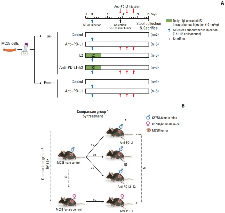
Fig. 1 Experimental design to evaluate the effects of anti–PD-L1 antibody and/or E2 on gut microbiome in the MC38 colon tumor mouse model. (A) Experimental design. A total of 5×10⁵ MC38 cells resuspended in 100 μL DPBS were injected subcutaneously into the right flank of 8-week-old C57BL/6 male mice. E2 (10 mg/kg) was intraperitoneally administered daily for 1 week from 3 days before injection of MC38 cells. Male and female mice bearing tumors (50–100 mm3) were selected and anti–PD-L1 or isotype control antibody was administered intraperitoneally at a dose of 10 mg/kg every 3 days for a total of three injections. Mice were sacrificed at day 26 after injection of MC38 cells. (B) Data analysis scheme. Investigation of changes in the gut microbiome composition according to treatment by comparing the gut microbiome in the MC38 males in the following groups: male control, anti–PD-L1–treated males, E2-treated males, males co-treated with anti–PD-L1 and E2. This analysis was used to examine the effect of anti–PD-L1 and/or E2 on the gut microbiome composition. Furthermore, sex-specific differences in the gut microbiome composition were analyzed by comparing the gut microbiome between male and female mice in the following groups: male control and female control, and anti–PD-L1 treated male and female groups. Based on this analysis, sex-specific changes in the gut microbiome composition were examined in MC38 colon tumor model. DPBS, Dulbecco’s phosphate-buffered saline; E2, 17β-estradiol; PD-L1, programmed death-ligand 1.
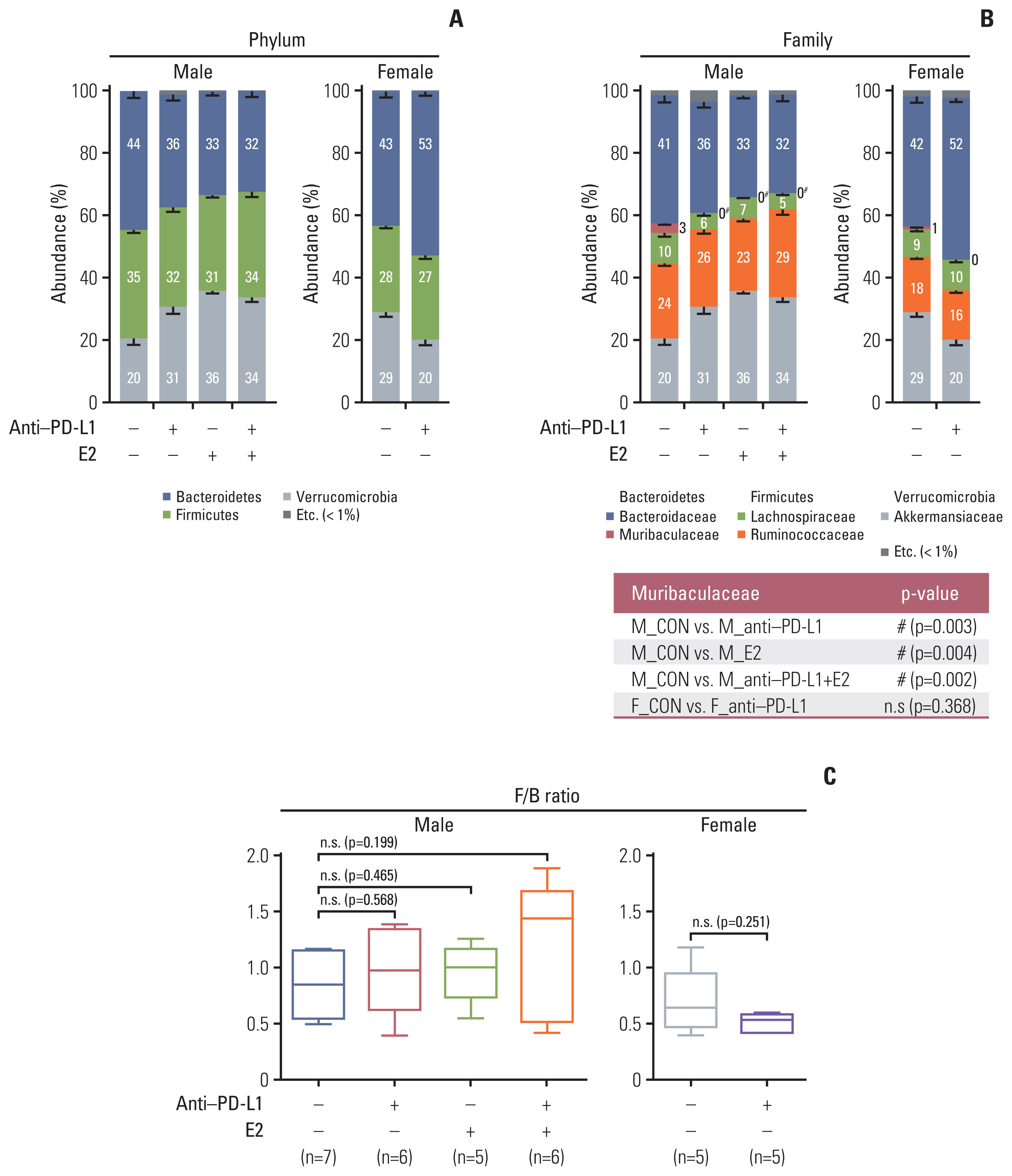
Fig. 2 Taxonomic composition. (A) Gut microbiota compositions at the phylum level in MC38 male (left) and female (right) groups. (B) Gut microbiota compositions at the family level in MC38 males (left) and females (right). Mann-Whitney U test was used for comparison of differences between two independent groups. The p-value for Muribaculaceae, which showed significance in the comparison between groups, is presented as a table under the figure. #p < 0.05, male control vs. anti–PD-L1–treated male, male control vs. E2-treated male, and male control vs. male co-treated with anti–PD-L1 and E2. (C) F/B ratio in MC38 males and females. Data are expressed as the mean±SEM. Whiskers show the minimum and maximum values. The p-values were calculated using the Mann-Whitney U test for comparison difference between independent two groups. E2, 17β-estradiol; F/B, Firmicutes/Bacteroidetes; n.s., not significant; PD-L1, programmed death-ligand 1; SEM, standard error of the mean.

Fig. 3 Beta diversity of gut microbiota. (A, B) Sample clustering using UniFrac-based PCoA and UPGMA tree at the species level in MC38 male groups. (C, D) Sample clustering using UniFrac-based PCoA and UPGMA tree at the species level in MC38 female groups. MC38 male and female samples were clustered using the Generalized UniFrac method at the species level. Significance of similarity of bacterial population structure was analyzed using PERMANOVA. The clustering and phylogenetic tree of each group are marked with a different color: M_CON, blue; M_anti-PD-L1, green; M_E2, grey; M_anti-PD-L1+E2, purple; F_CON, red; F_anti-PD-L1, orange color. CON, control; E2, 17β-estradiol; F, female; M, male; PCoA, principal coordinates analysis; PD-L1, programmed death-ligand 1; PERMANOMA, permutational multivariate analysis of variance; UPGMA, unweighted pair group method with arithmetic mean.

Fig. 4 Identification of specific gut microbiome for anti–PD-L1 and/or E2 in MC38 colon tumor model. (A–D) LEfSe analysis. Bar plots of the LEfSe results, which were obtained based on the following criteria: (1) alpha value for the factorial Kruskal-Wallis test among classes < 0.05; (2) the alpha value for the pairwise Wilcoxon test between subclasses < 0.05; (3) threshold on the logarithmic LDA score for discriminative features < 2.0; and (4) a multi-class analysis set as all-against-all. Bacterial characteristics were classified as “commensal bacteria,” “opportunistic pathogens” and “uncharacterized” according to previous reports. The color bars show the LDA score (log10) of species that enriched in indicated conditions; (A–C) blue bar (male control), (A) block bar (anti–PD-L1–treated male), (B) green bar (E2-treated male), (C) purple bar (male co-treated with anti–PD-L1 and E2), (D) red bar (female control), gray bar (anti–PD-L1–treated female). The color on the species name indicates the characteristics of each species: yellow for commensal bacteria, orange for opportunistic pathogens, and no color for not characterized bacteria. (E, F) Venn diagrams based on LEfSe data. (E) Identification of bacteria exhibiting changes after anti–PD-L1 and/or E2 treatment in MC38 male mice. (F) Identification of bacteria exhibiting common changes after anti–PD-L1 treatment in MC38 male and female mice. CON, control; E2, 17β-estradiol; F, female; LDA, linear discriminant analysis; LEfSe, LDA effect size; M, male; PD-L1, programmed death-ligand 1.
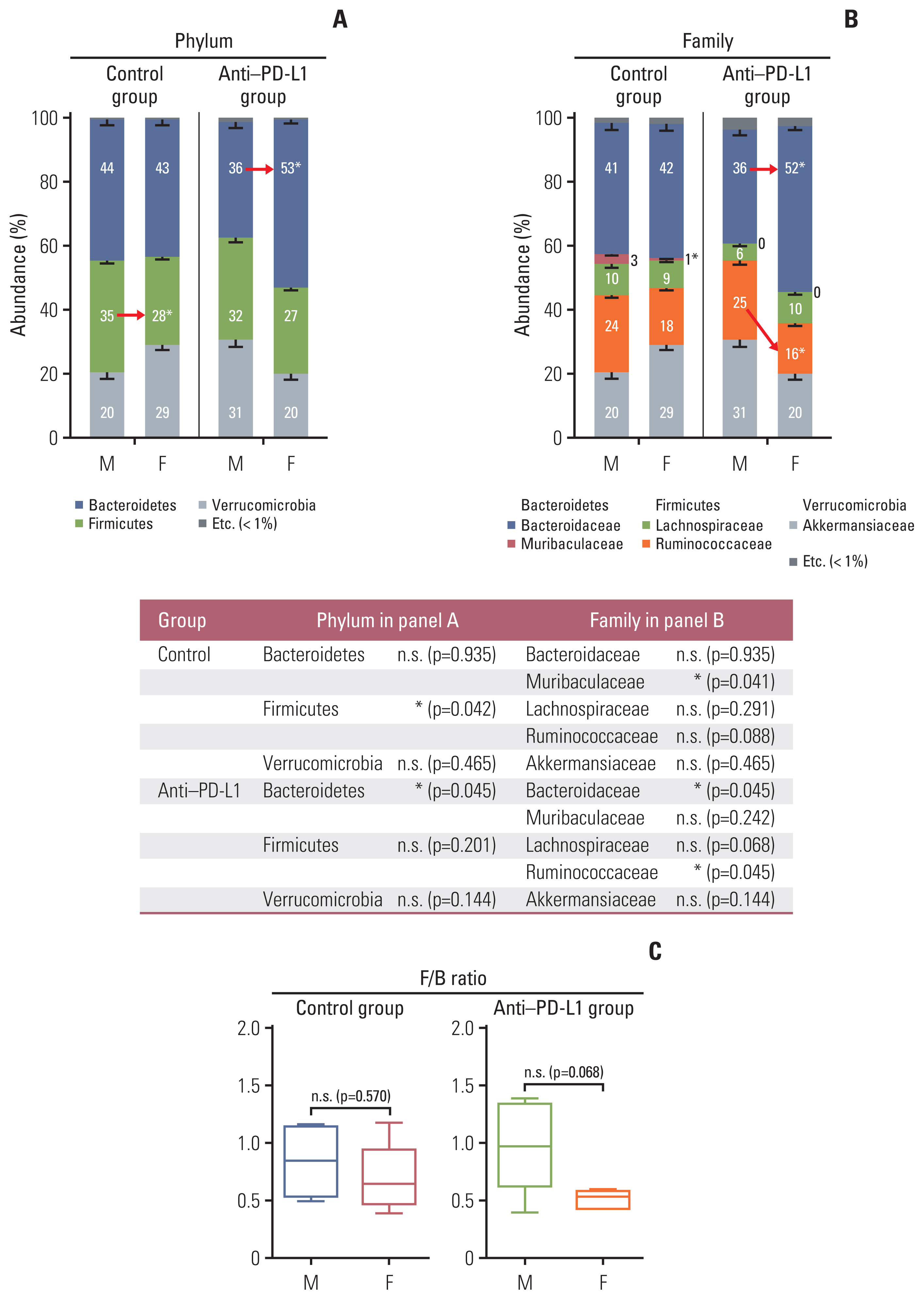
Fig. 5 Taxonomic composition. (A) Gut microbiota compositions at the phylum level in control group (left) and anti–PD-L1 group (right) in MC38 male and female mice. (B) Gut microbiota compositions at the family level in control group (left) and anti–PD-L1 group (right) in MC38 male and female mice. Mann-Whitney U test was used for comparison of differences between two independent groups. The p-values for Phylum and Family level are presented as a table under the figure. *p < 0.05, male control vs female control, anti–PD-L1–treated male vs. anti–PD-L1–treated female. (C) F/B ratio in control groups and anti–PD-L1 treated groups in MC38 male and female mice. Data are expressed as the mean±SEM. Whiskers show the minimum and maximum values. The p-values were calculated using the Mann-Whitney U test for comparison of differences between two independent groups. F, female; F/B, Firmicutes/Bacteroidetes; M, male; n.s., not significant; PD-L1, programmed death-ligand 1; SEM, standard error of the mean.
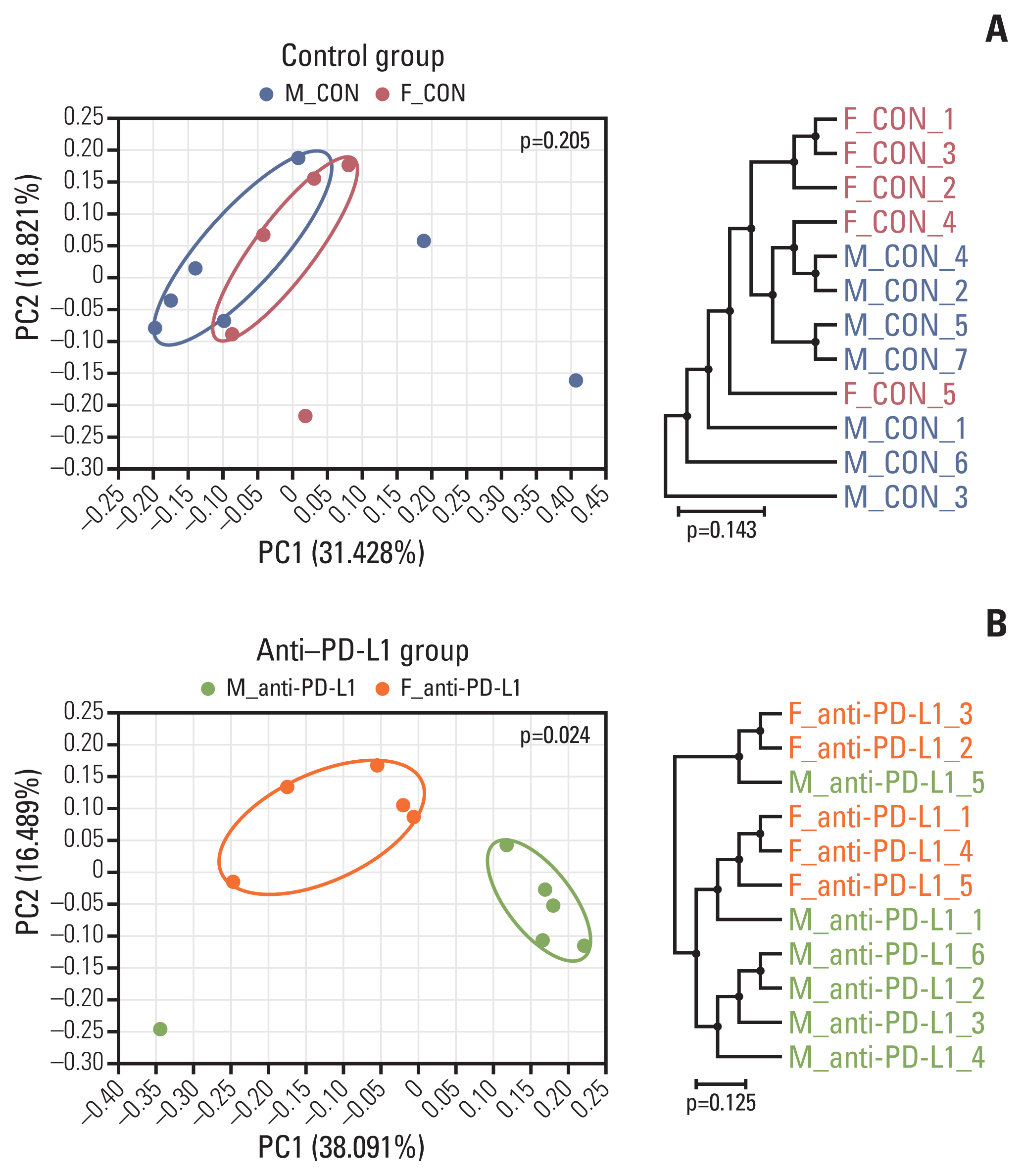
Fig. 6 Beta diversity of gut microbiota. (A) Sample clustering using UniFrac-based PCoA and UPGMA tree at the species level in MC38 male and female control groups. (B) Sample clustering using UniFrac-based PCoA and UPGMA tree at the species level in anti–PD-L1–treated MC38 male and female groups. MC38 male and female samples were clustered using the Generalized UniFrac method at the species level. Significance for similarity of bacterial population structure was analyzed using PERMANOVA. The clustering and phylogenetic tree of each group are marked with a different color: M_CON, blue; F_CON, red; M_anti-PD-L1, green; F_anti-PD-L1, orange color. CON, control; F, female; M, male; PCoA, principal coordinates analysis; PD-L1, programmed death-ligand 1; PERMANOMA, permutational multivariate analysis of variance; UPGMA, unweighted pair group method with arithmetic mean.

Fig. 7 Identification of sex-specific gut microbiome in control and anti–PD-L1 group in MC38 male and female mice. (A, B) LEfSe analysis. Bar plots of the LEfSe results, which were obtained based on the same criteria mentioned in Fig. 5. The color bars show the LDA score (log10) of species that were enriched in indicated conditions; (A) blue bar (male control) and red bar (female control), (B) block bar (anti–PD-L1–treated male) and gray bar (anti–PD-L1-treated female). (C) Venn diagrams based on LEfSe data. Identification of bacteria showing sex-dependent changes in control and anti–PD-L1 groups in male and female MC38 mice. CON, control; E2, 17β-estradiol; F, female; LDA, linear discriminant analysis; LEfSe, LDA effect size; M, male; PD-L1, programmed death-ligand 1.

Fig. 8 Regulatory mechanism by which estrogen modulates the tumor microenvironment and simultaneously alters the gut microbiome to increase the effect of anti–PD-L1 and contribute to tumor size reduction in the MC38 colon tumor model. CAFs, cancer-associated fibroblasts; E2, 17β-estradiol; PD-1, programmed cell death-1; PD-L1, programmed death-ligand 1; TAM, tumor-associated macrophages.
Reference
-
References
1. Shanahan F. The colonic microbiota in health and disease. Curr Opin Gastroenterol. 2013; 29:49–54.
Article2. Ternes D, Karta J, Tsenkova M, Wilmes P, Haan S, Letellier E. Microbiome in colorectal cancer: how to get from meta-omics to mechanism? Trends Microbiol. 2020; 28:401–23.
Article3. Chen Y, Chen Y, Zhang J, Cao P, Su W, Deng Y, et al. Fusobacterium nucleatum promotes metastasis in colorectal cancer by activating autophagy signaling via the upregulation of CARD3 expression. Theranostics. 2020; 10:323–39.
Article4. Zhang S, Yang Y, Weng W, Guo B, Cai G, Ma Y, et al. Fusobacterium nucleatum promotes chemoresistance to 5-fluorouracil by upregulation of BIRC3 expression in colorectal cancer. J Exp Clin Cancer Res. 2019; 38:14.
Article5. Kim SE, Paik HY, Yoon H, Lee JE, Kim N, Sung MK. Sex- and gender-specific disparities in colorectal cancer risk. World J Gastroenterol. 2015; 21:5167–75.
Article6. Chlebowski RT, Wactawski-Wende J, Ritenbaugh C, Hubbell FA, Ascensao J, Rodabough RJ, et al. Estrogen plus progestin and colorectal cancer in postmenopausal women. N Engl J Med. 2004; 350:991–1004.
Article7. Baker JM, Al-Nakkash L, Herbst-Kralovetz MM. Estrogen-gut microbiome axis: physiological and clinical implications. Maturitas. 2017; 103:45–53.
Article8. Chen KL, Madak-Erdogan Z. Estrogen and microbiota crosstalk: should we pay attention? Trends Endocrinol Metab. 2016; 27:752–5.
Article9. Org E, Mehrabian M, Parks BW, Shipkova P, Liu X, Drake TA, et al. Sex differences and hormonal effects on gut microbiota composition in mice. Gut Microbes. 2016; 7:313–22.
Article10. Song CH, Kim N, Nam RH, Choi SI, Lee HN, Surh YJ. 17beta-Estradiol supplementation changes gut microbiota diversity in intact and colorectal cancer-induced ICR male mice. Sci Rep. 2020; 10:12283.11. Cox-York KA, Sheflin AM, Foster MT, Gentile CL, Kahl A, Koch LG, et al. Ovariectomy results in differential shifts in gut microbiota in low versus high aerobic capacity rats. Physiol Rep. 2015; 3:e12488.
Article12. Marin-Acevedo JA, Kimbrough EO, Lou Y. Next generation of immune checkpoint inhibitors and beyond. J Hematol Oncol. 2021; 14:45.
Article13. Robert C. A decade of immune-checkpoint inhibitors in cancer therapy. Nat Commun. 2020; 11:3801.
Article14. Wei SC, Duffy CR, Allison JP. Fundamental mechanisms of immune checkpoint blockade therapy. Cancer Discov. 2018; 8:1069–86.15. Son HJ, Sohn SH, Kim N, Lee HN, Lee SM, Nam RH, et al. Effect of estradiol in an azoxymethane/dextran sulfate sodium-treated mouse model of colorectal cancer: implication for sex difference in colorectal cancer development. Cancer Res Treat. 2019; 51:632–48.
Article16. Song CH, Kim N, Nam RH, Choi SI, Jang JY, Kim JW, et al. Combination treatment with 17beta-estradiol and anti-PD-L1 suppresses MC38 tumor growth by reducing PD-L1 expression and enhancing M1 macrophage population in MC38 colon tumor model. Cancer Lett. 2022; 543:215780.17. Hou W, Sampath P, Rojas JJ, Thorne SH. Oncolytic virus-mediated targeting of PGE2 in the tumor alters the immune status and sensitizes established and resistant tumors to immunotherapy. Cancer Cell. 2016; 30:108–19.
Article18. Shi G, Yang Q, Zhang Y, Jiang Q, Lin Y, Yang S, et al. Modulating the tumor microenvironment via oncolytic viruses and CSF-1R inhibition synergistically enhances anti-PD-1 immunotherapy. Mol Ther. 2019; 27:244–60.19. Lee CH, Bae JH, Choe EJ, Park JM, Park SS, Cho HJ, et al. Macitentan improves antitumor immune responses by inhibiting the secretion of tumor-derived extracellular vesicle PD-L1. Theranostics. 2022; 12:1971–87.
Article20. Karp NA, Wilson Z, Stalker E, Mooney L, Lazic SE, Zhang B, et al. A multi-batch design to deliver robust estimates of efficacy and reduce animal use - a syngeneic tumour case study. Sci Rep. 2020; 10:6178.
Article21. Bolger AM, Lohse M, Usadel B. Trimmomatic: a flexible trimmer for Illumina sequence data. Bioinformatics. 2014; 30:2114–20.
Article22. Rognes T, Flouri T, Nichols B, Quince C, Mahe F. VSEARCH: a versatile open source tool for metagenomics. PeerJ. 2016; 4:e2584.
Article23. Myers EW, Miller W. Optimal alignments in linear space. Comput Appl Biosci. 1988; 4:11–7.
Article24. Wheeler TJ, Eddy SR. nhmmer: DNA homology search with profile HMMs. Bioinformatics. 2013; 29:2487–9.
Article25. Yoon SH, Ha SM, Kwon S, Lim J, Kim Y, Seo H, et al. Introducing EzBioCloud: a taxonomically united database of 16S rRNA gene sequences and whole-genome assemblies. Int J Syst Evol Microbiol. 2017; 67:1613–7.
Article26. Edgar RC, Haas BJ, Clemente JC, Quince C, Knight R. UCH-IME improves sensitivity and speed of chimera detection. Bioinformatics. 2011; 27:2194–200.
Article27. Segata N, Izard J, Waldron L, Gevers D, Miropolsky L, Garrett WS, et al. Metagenomic biomarker discovery and explanation. Genome Biol. 2011; 12:R60.
Article28. Wu TR, Lin CS, Chang CJ, Lin TL, Martel J, Ko YF, et al. Gut commensal Parabacteroides goldsteinii plays a predominant role in the anti-obesity effects of polysaccharides isolated from Hirsutella sinensis. Gut. 2019; 68:248–62.
Article29. Lai CH, Lin TL, Huang MZ, Li SW, Wu HY, Chiu YF, et al. Gut commensal Parabacteroides goldsteinii MTS01 alters gut microbiota composition and reduces cholesterol to mitigate Helicobacter pylori-induced pathogenesis. Front Immunol. 2022; 13:916848.
Article30. Ervin SM, Li H, Lim L, Roberts LR, Liang X, Mani S, et al. Gut microbial beta-glucuronidases reactivate estrogens as components of the estrobolome that reactivate estrogens. J Biol Chem. 2019; 294:18586–99.31. Tang C, Kamiya T, Liu Y, Kadoki M, Kakuta S, Oshima K, et al. Inhibition of dectin-1 signaling ameliorates colitis by inducing Lactobacillus-mediated regulatory T cell expansion in the intestine. Cell Host Microbe. 2015; 18:183–97.
Article32. Wilck N, Matus MG, Kearney SM, Olesen SW, Forslund K, Bartolomaeus H, et al. Salt-responsive gut commensal modulates T(H)17 axis and disease. Nature. 2017; 551:585–9.
Article33. Song CH, Kim N, Nam RH, Choi SI, Yu JE, Nho H, et al. Changes in microbial community composition related to sex and colon cancer by Nrf2 knockout. Front Cell Infect Microbiol. 2021; 11:636808.
Article34. Lee SH, Cho SY, Yoon Y, Park C, Sohn J, Jeong JJ, et al. Bifidobacterium bifidum strains synergize with immune checkpoint inhibitors to reduce tumour burden in mice. Nat Microbiol. 2021; 6:277–88.
Article
- Full Text Links
-
- Actions
-
Cited
- CITED
-
- Close
- Share
-
- Similar articles
-
- The Expression of Programmed Death-Ligand 1 on Immune Cells Is Related to a Better Prognosis in Biliary Tract Cancer
- PD-1: A Negative Regulator of Phagocytosis by Tumour-Associated Macrophages in Colon Cancer
- A Novel Anti-PD-L1 Antibody Exhibits Antitumor Effects on Multiple Myeloma in Murine Models via Antibody-Dependent Cellular Cytotoxicity
- Peripheral blood immune cell-based biomarkers in anti-PD-1/PD-L1 therapy
- Characteristics of Immune-Related Thyroid Adverse Events in Patients Treated with PD-1/PD-L1 Inhibitors
