Noradrenergic axons hitch hiking along the human abducens nerve
- Affiliations
-
- 1Department of Otolaryngology, Henry Ford Macomb Hospital, Detroit, MI, USA
- 2Department of Anatomy, Lake Erie College of Osteopathic Medicine, Erie, PA, USA
- KMID: 2544090
- DOI: http://doi.org/10.5115/acb.22.223
Abstract
- The abducens nerve (AN; cranial nerve VI) exits the brainstem at the inferior pontine sulcus, pierces the dura of the posterior cranial fossa, passes through the cavernous sinus in close contact to the internal carotid artery (ICA) and traverses the superior orbital fissure to reach the orbit to innervate the lateral rectus muscle. At its exit from the brainstem, the AN includes only axons from lower motor neurons in the abducens nucleus. However, as the AN crosses the ICA it receives a number of branches from the internal carotid sympathetic plexus. The arrangement, neurochemical profile and function of these sympathetic axons running along the AN remain unresolved. Herein, we use gross dissection and microscopic study of hematoxylin and eosin-stained sections and sections with tyrosine hydroxylase immunolabeling. Our results suggest the AN receives multiple bundles of unmyelinated axons that use norepinephrine as a neurotransmitter consistent with postganglionic sympathetic axons.
Figure
-
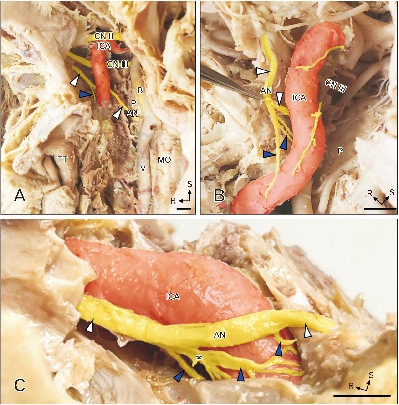
Fig. 1 Contribution from the ICP to the AN. (A, B) are different stages of the dissection of the right ICA and cavernous sinus of a single specimen. (A) The ICA has been exposed from the anterior aspect and the cavernous sinus has been dissected away. The optic nerve (CN II), oculomotor nerve (CN III) and AN are visible. The AN (white arrowheads) can be seen emerging from the brainstem and coursing lateral to the ICA. Branches from the ICP joining the AN are indicated by blue arrowheads. A deeper dissection of this region is (B) the AN (white arrowheads) is pulled away from the ICA with forceps to demonstrate the branches from the ICP (blue arrowheads). (C) is a dissection of the ICA and cavernous sinus from the left side of a different specimen where the venous plexus, CN II and branches for CN V have been removed. The AN (white arrowheads) courses lateral to the ICA and is joined by multiple branches from the ICP (blue arrowheads). The scale bars are all equal to 5 mm. ICP, internal carotid sympathetic plexus; AN, abducens nerve; ICA, internal carotid artery; TT, torus tubaris, V, vertebral artery; MO, medulla oblongata; P, pons; B, basilar artery; R, rostral; S, superior.
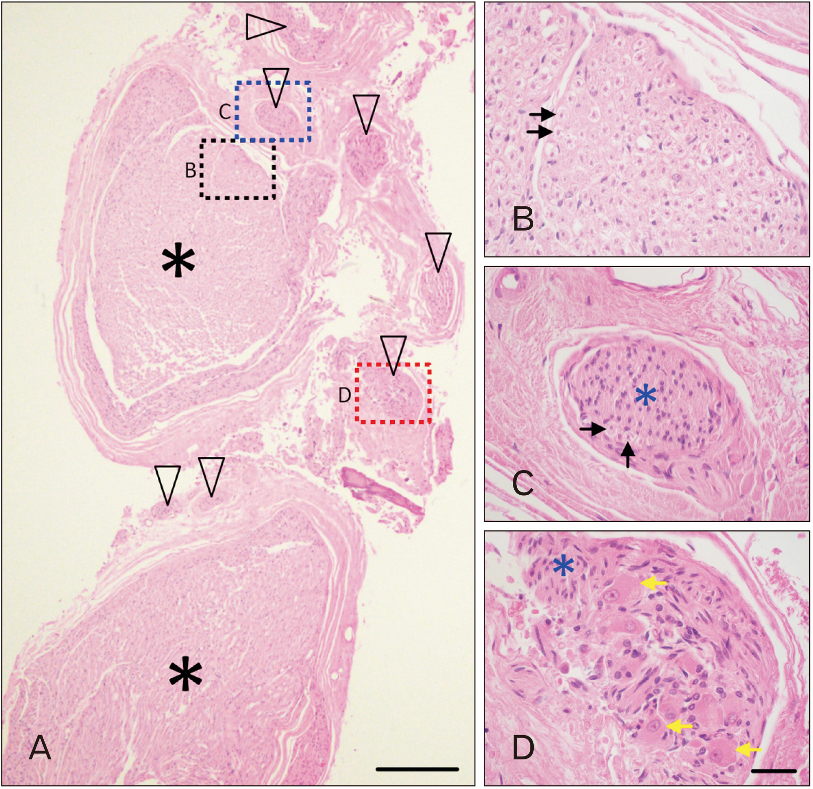
Fig. 2 Unmyelinated axons along the AN. (A) is a low magnification image of an H&E-stained section through the cavernous segment of the AN. The fascicles indicated by black asterisks are composed of myelinated axons; the fascicles indicated by the black arrowheads are largely composed of unmyelinated axons. (B) shows a higher magnification view of the region indicated by the black dashed box. Individual myelinated axons are indicated by the black arrows. (C) shows a higher magnification view of the region indicated by the blue dashed box. This bundle of axons (blue asterisk) contains mainly unmyelinated axons; myelinated axons are indicated by the black arrows. (D) shows a higher magnification view of the region indicated by the red dashed box. This fascicle contains only unmyelinated axons and a collection of neuronal cell bodies (yellow arrows). The scale bar in A is equal to 300 mm; the scale bar in D is equal to 40 mm and applies to B–D. AN, abducens nerve.
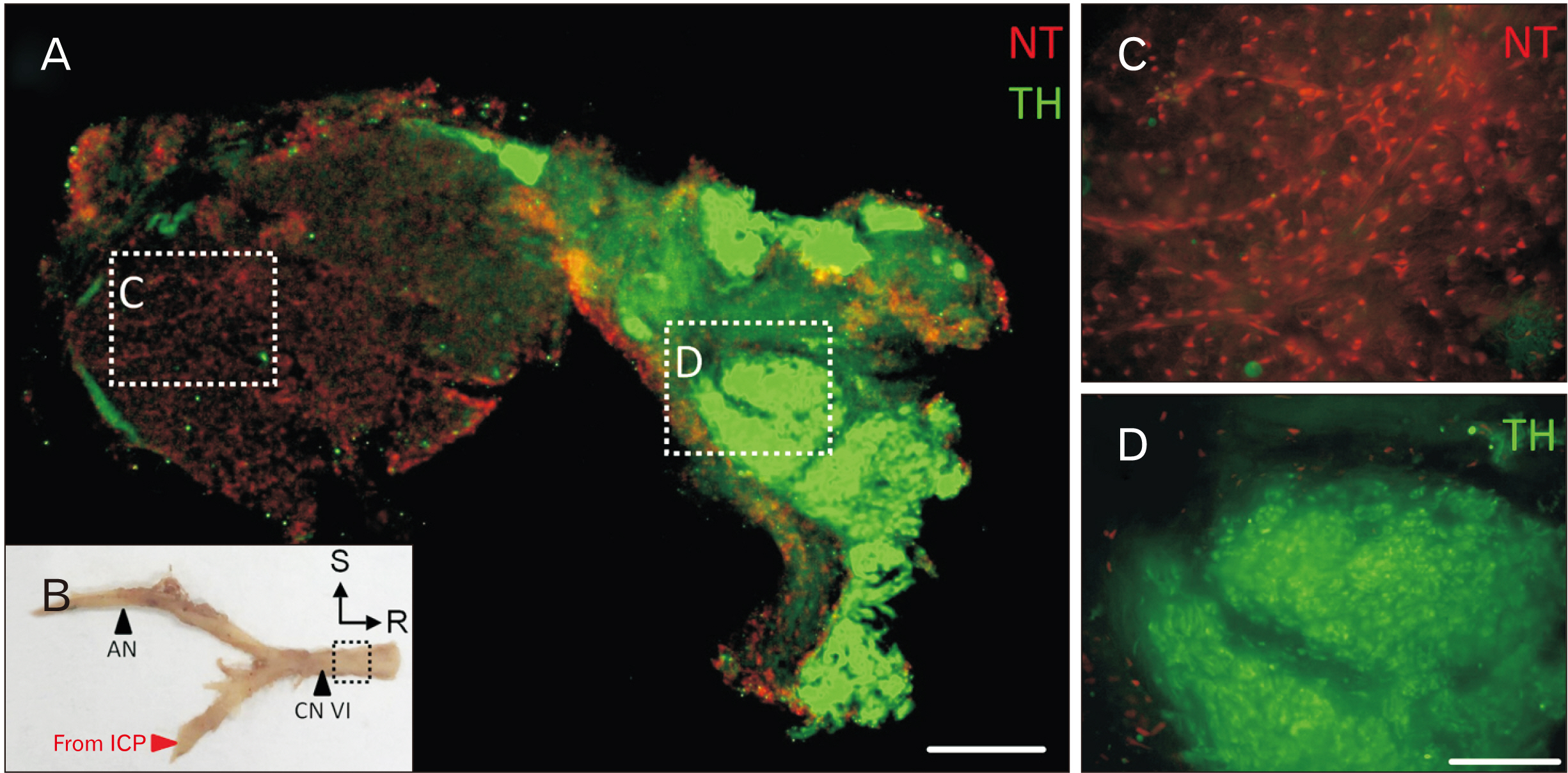
Fig. 3 TH+ axons along the AN. (A) shows a cross section of the AN labeled for TH and counterstained with neurotrace red (NT). This section is taken from the AN distal to receiving branches from the ICP (boxed area in B). A higher magnification view of the main trunk of the AN is (C); there is no TH immunolabeling in this part of the nerve. A higher magnification view of the peripheral aspect of the nerve is (D); the majority of these axons are TH+, consistent with noradrenergic function. The scale bar in A is equal to 200 mm; the scale bar in D is equal to 40 mm and applies to both C and D. AN, abducens nerve; S, superior; R, rostral; ICP, internal carotid sympathetic plexus.
Reference
-
References
1. Standring S. 2016. Gray's anatomy: the anatomical basis of clinical practice. 41st ed. Elsevier;DOI: 10.1002/ca.22677.2. Cunningham DJ, Robinson A. 1931. Cunningham's text-book of anatomy. 6th ed. Oxford University Press;DOI: 10.5962/bhl.title.44024.3. Monro A. 1746. The anatomy of the human bones and nerves. 4th ed. Hamilton and Balfour;DOI: 10.5962/bhl.title.114861.4. Johnston JA, Parkinson D. 1974; Intracranial sympathetic pathways associated with the sixth cranial nerve. J Neurosurg. 40:236–43. DOI: 10.3171/jns.1974.40.2.0236. PMID: 4809122.
Article5. Quain J, Leidy J, Quain R, Sharpey W. 1849. Human anatomy. 1st ed. Vol 2:Lea and Blanchard;p. 343–4.6. Gray H, Pick TP, Howden R. 1901. Anatomy, descriptive and surgical. 15th ed. Lea Brothers;p. 802. DOI: 10.5962/bhl.title.31366.7. Plarr V, Power D, Spencer WG, Gask GE. 1930. Plarr's lives of the fellows of the Royal College of Surgeons of England. Vol 1:John Wright & Sons;p. 464.8. McGrath P. 1977; The cavernous sinus: an anatomical survey. Aust N Z J Surg. 47:601–13. DOI: 10.1111/j.1445-2197.1977.tb06591.x. PMID: 273404.
Article9. Parkinson D, Johnston J, Chaudhuri A. 1978; Sympathetic connections to the fifth and sixth cranial nerves. Anat Rec. 191:221–6. DOI: 10.1002/ar.1091910207. PMID: 666018.
Article10. Bleys RL, Janssen LM, Groen GJ. 2001; The lateral sellar nerve plexus and its connections in humans. J Neurosurg. 95:102–10. DOI: 10.3171/jns.2001.95.1.0102. PMID: 11453377.
Article11. Iwanaga J, Anand MK, Camacho A, Rodriguez F, Watson C, Caskey EL, Dumont AS, Tubbs RS. 2020; Surgical anatomy of the internal carotid plexus branches to the abducens nerve in the cavernous sinus. Clin Neurol Neurosurg. 191:105690. DOI: 10.1016/j.clineuro.2020.105690. PMID: 31982693.
Article12. Wysiadecki G, Radek M, Tubbs RS, Iwanaga J, Walocha J, Brzeziński P, Polguj M. 2021; Gross and micro-anatomical study of the cavernous segment of the abducens nerve and its relationships to internal carotid plexus: application to skull base surgery. Brain Sci. 11:649. DOI: 10.3390/brainsci11050649. PMID: 34065668. PMCID: PMC8156379. PMID: 7c47d66c3189421a831c3e0d82f96484.
Article13. Pickel VM, Joh TH, Reis DJ. 1975; Ultrastructural localization of tyrosine hydroxylase in noradrenergic neurons of brain. Proc Natl Acad Sci U S A. 72:659–63. DOI: 10.1073/pnas.72.2.659. PMID: 235760. PMCID: PMC432374.
Article14. Gellért A. 1934; Ganglia of the internal carotid plexus. J Anat. 68(Pt 3):318–22. PMID: 17104480. PMCID: PMC1249031.15. Carvalho VC. 1985; Nerve cells in the human cavernous sinus. Anat Anz. 159:29–32. PMID: 4096407.
- Full Text Links
-
- Actions
-
Cited
- CITED
-
- Close
- Share
-
- Similar articles
-
- A Case of Isolated Unilateral Abducens Nerve Palsy Caused by Clival Metastasis from Rectal Cancer
- Unilateral Abducens Nerve Palsy Associated with Ruptured Anterior Communicating Artery Aneurysm
- Cystic Abducens Schwannoma without Abducens Paresis : Possible Role of Cisternal Structures in Clinical Manifestation
- A Case of Traumatic Bilateral Abducens Nerve Palsy Associated with Skull Base Fracture
- A Patient Presented With Unilateral Abducens Nerve Palsy: A Variant Form of Guillain-Barre Syndrome With Anti-GT1a Antibody
