Development and growth of the human fetal sacroiliac joint revisited: a comparison with the temporomandibular joint
- Affiliations
-
- 1Department of Anatomy, Jeonbuk National University Medical School, Jeonju, Korea
- 2Department of Anatomy, Wuxi School of Medicine, Jiangnan University, Wuxi, Jiangsu, China
- 3Department of Anatomy, Tokai University School of Medicine, Isehara, Japan
- 4Division of Internal Medicine, Cupid Clinic, Iwamizawa, Japan
- 5Emeritus professor of Akita University School of Medicine, Akita, Japan
- 6Department of Anatomy and Human Embryology, Institute of Embryology, Complutense University, Madrid, Spain
- KMID: 2544087
- DOI: http://doi.org/10.5115/acb.22.189
Abstract
- The human fetal sacroiliac joint (SIJ) is characterized by unequal development of the paired bones and delayed cavitation. Thus, during the long in utero period, the bony ilium becomes adjacent to the cartilaginous sacrum. This mor phology may be analogous to that of the temporomandibular joint (TMJ). We examined horizontal histological sections of 24 fetuses at 10–30 weeks and compared the timing and sequences of joint cartilage development, cavitation, and ossification of the ilium. We also examined histological sections of the TMJ and humeroradial joint, because these also contain a disk or disk-like structure. In the ilium, endochondral ossification started in the anterior side of the SIJ, extended posteriorly and reached the joint at 12 weeks GA, and then extended over the joint at 15 weeks GA. Likewise, the joint cartilage appeared at the anterior end of the future SIJ at 12 weeks GA, and extended along the bony ilium posteriorly to cover the entire SIJ at 26 weeks GA. The cavitation started at 15 weeks GA. Therefore, joint cartilage development seemed to follow the ossification of the ilium by extending along the SIJ, and cavitation then occurred. This sequence “ossification, followed by joint cartilage formation, and then cavitation” did not occur in the TMJ or humeroradial joint. The TMJ had a periosteum-like membrane that covered the joint surface, but the humeroradial joint did not. After muscle contraction starts, it is likely that the mechanical stress from the bony ilium induces development of joint cartilage.
Figure
-

Fig. 1 Horizontal sections of sacroiliac joints in four fetuses (crown-rump length: 52–90 mm) that were stained with H&E (A) or azan (B–D). (A, B) show that ossification of the ilium occurs in the anterior part, but does not reach the future joint area. (A) shows membranous bones (arrowheads), not cartilaginous bones. (C) shows a small cartilage mass attached to the ilium (arrow). (D) shows that the cartilage covers almost one-third of the future joint area (arrow). The circles in (B–D) indicate endochondral ossification of the ilium. The asterisks in (A, D) indicate an artifact space produced during histological procedure. (A–D) Scale bars=1 mm.

Fig. 2 Horizontal sections of sacroiliac joints in four fetuses (crown-rump length: 120–232 mm) that were stained with H&E (A, B, D) or Masson’s trichrome (C). All panels show the bony ilium covering the joint area. (A–C) show that the joint cartilage of the ilium (arrows) covers the anterior half of the joint area. (D) shows the cartilage extends posteriorly over the joint. (B, D) show the joint cavities (stars). The insert on the left of (C) (corresponding to the rectangle in the main panel) shows CD68-positive cells at higher magnification, in which the future joint space (triangles) contains no macrophages. The circles in (A, C) indicate endochondral ossification of the ilium. The asterisk in (A) indicates an artifact space produced during the histological procedure. All panels (except the C insert) were at the same magnification. (A) Scale bar=1 mm.

Fig. 3 Sagittal sections of temporomandibular joints in three fetuses (crown- rump length: 125, 210, and 250 mm) that were stained with H&E. (A) shows attachment of the condylar cartilage to the bony temporal bone, with initiation of ossification in the condyle (arrowheads) and cavitation (stars). (B, C) show that the joint cavity is large and the disk is thick, but a periosteum-like membrane (arrows) covers the joint surface of the condyle and temporal bone, instead of the joint cartilage. Ovals in (C) show endochondral ossification occurs in the condyle. (A–C) Scale bars=1 mm.
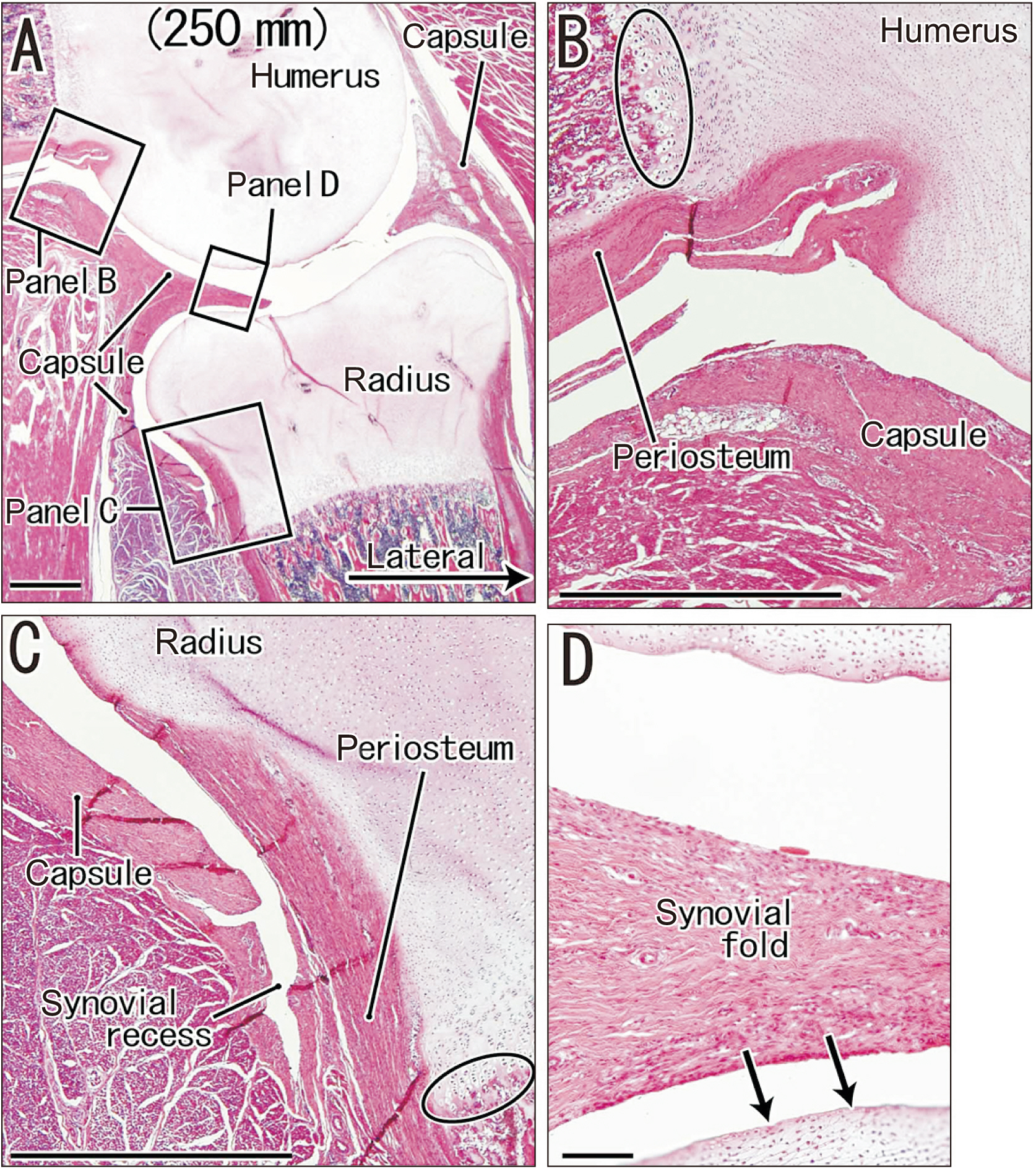
Fig. 4 Sagittal sections of the humeroradial joint in a fetus (crown-rump length: 250 mm) that were stained with H&E. (A) shows the topographical anatomy around the humeroradial joint. (B–D) are higher magnification views of three rectangles in (A). (B, C) show the periosteum and joint capsule at recesses of the joint cavity. (D) shows a thick synovial fold and its adjacent joint cartilage, and that the periosteum does not continue to the joint surface in the humeroradial joint (arrows). The oval in (B) shows the endochondral ossification is at the distal end of the periosteum in the humerus, and the oval in (C) shows it is covered by the periosteum in the radius. (A) Scale bar=1 mm; (B–D) Scale bars=0.1 mm.
Reference
-
References
1. Schunke GB. 1938; The anatomy and development of the sacro-iliac joint in man. Anat Rec. 72:313–31. DOI: 10.1002/ar.1090720306.
Article2. Vleeming A, Schuenke MD, Masi AT, Carreiro JE, Danneels L, Willard FH. 2012; The sacroiliac joint: an overview of its anatomy, function and potential clinical implications. J Anat. 221:537–67. DOI: 10.1111/j.1469-7580.2012.01564.x. PMID: 22994881. PMCID: PMC3512279.
Article3. Shibata S, Sato R, Murakami G, Fukuoka H, Rodríguez-Vázquez JF. 2013a; Origin of mandibular condylar cartilage in mice, rats, and humans: periosteum or separate blastema? J Oral Biosci. 55:208–16. DOI: 10.1016/j.job.2013.08.001.
Article4. Shibata S, Sakamoto Y, Baba O, Qin C, Murakami G, Cho BH. 2013b; An immunohistochemical study of matrix proteins in the craniofacial cartilage in midterm human fetuses. Eur J Histochem. 57:e39. DOI: 10.4081/ejh.2013.e39. PMID: 24441192. PMCID: PMC3896041. PMID: 810700d8add242cea1a99dda671f53e3.
Article5. Shibata S, Sakamoto Y, Yokohama-Tamaki T, Murakami G, Cho BH. 2014; Distribution of matrix proteins in perichondrium and periosteum during the incorporation of Meckel's cartilage into ossifying mandible in midterm human fetuses: an immunohistochemical study. Anat Rec (Hoboken). 297:1208–17. DOI: 10.1002/ar.22911. PMID: 24700703.
Article6. Mérida-Velasco JA, Sánchez-Montesinos I, Espín-Ferra J, Rodríguez-Vázquez JF, Mérida-Velasco JR, Jiménez-Collado J. 1997; Development of the human knee joint. Anat Rec. 248:269–78. DOI: 10.1002/(SICI)1097-0185(199706)248:2<269::AID-AR14>3.0.CO;2-N. PMID: 9185993.
Article7. GARDNER E, GRAY DJ. 1950; Prenatal development of the human hip joint. Am J Anat. 87:163–211. DOI: 10.1002/aja.1000870202. PMID: 14771010.
Article8. Bowen V, Cassidy JD. 1981; Macroscopic and microscopic anatomy of the sacroiliac joint from embryonic life until the eighth decade. Spine (Phila Pa 1976). 6:620–8. DOI: 10.1097/00007632-198111000-00015. PMID: 7336283.
Article9. Ishimine T. 1989; Histopathological study of the aging process in the human sacroiliac joint. Nihon Seikeigeka Gakkai Zasshi. 63:1074–84. Japanese. PMID: 2584838.10. Uhthoff HK. 1993; Prenatal development of the iliolumbar ligament. J Bone Joint Surg Br. 75:93–5. DOI: 10.1302/0301-620X.75B1.8421046. PMID: 8421046.
Article11. Bogduk N. Bogduk N, editor. 1997. The sacroiliac joint. Clinical Anatomy of the Lumbar Spine and Sacrum. 3rd ed. Churchill Livingstone;New York: p. 177–85.12. Kampen WU, Tillmann B. 1998; Age-related changes in the articular cartilage of human sacroiliac joint. Anat Embryol (Berl). 198:505–13. DOI: 10.1007/s004290050200. PMID: 9833689.
Article13. Ikeno H, Matsumura H, Murakami G, Sato TJ, Ohta M. 2006; Which morphology of dry bone articular surfaces suggests so-called fibrous ankylosis in the elderly human sacroiliac joint? Anat Sci Int. 81:39–46. DOI: 10.1111/j.1447-073X.2006.00126.x. PMID: 16526595.
Article14. Isogai S, Murakami G, Wada T, Ishii S. 2001; Which morphologies of synovial folds result from degeneration and/or aging of the radiohumeral joint: an anatomic study with cadavers and embryos. J Shoulder Elbow Surg. 10:169–81. DOI: 10.1067/mse.2001.112956. PMID: 11307082.
Article15. Naito T, Cho KH, Yamamoto M, Hirouchi H, Murakami G, Hayashi S, Abe S. 2019; Examination of the topographical anatomy and fetal development of the tendinous annulus of Zinn for a common origin of the extraocular recti. Invest Ophthalmol Vis Sci. 60:4564–73. DOI: 10.1167/iovs.19-28094. PMID: 31675425.
Article16. Yamamoto M, Jin ZW, Hayashi S, Rodríguez-Vázquez JF, Murakami G, Abe S. 2021; Association between the developing sphenoid and adult morphology: a study using sagittal sections of the skull base from human embryos and fetuses. J Anat. 239:1300–17. DOI: 10.1111/joa.13515. PMID: 34268732.
Article17. Jin ZW, Jin Y, Yamamoto M, Abe H, Murakami G, Yan TF. 2016; Oblique cord (chorda obliqua) of the forearm and muscle-associated fibrous tissues at and around the elbow joint: a study of human foetal specimens. Folia Morphol (Warsz). 75:493–502. DOI: 10.5603/FM.a2016.0019. PMID: 27830875.
Article18. Abe H, Hayashi S, Kim JH, Murakami G, Rodríguez-Vázquez JF, Jin ZW. 2021; Fetal development of the thoracolumbar fascia with special reference to the fascial connection with the transversus abdominis, latissimus dorsi, and serratus posterior inferior muscles. Surg Radiol Anat. 43:917–28. DOI: 10.1007/s00276-020-02668-4. PMID: 33438110.
Article19. Sato T, Kim JH, Cho KH, Hayashi S, Rodríguez-Vázquez JF, Murakami G. 2021; Fetal development and growth of the human erector spinae with special reference to attachments on the surface aponeurosis. Surg Radiol Anat. 43:1503–17. DOI: 10.1007/s00276-021-02759-w. PMID: 34059927.
Article20. Moffet BC. 1957; The prenatal development of the human temporomandibular joint. Contr Embryo. 36:19–28.21. Xiang L, Wang X, Li Y, Liu HW, Zhang X, Mu X, Liu C, Hu M. 2022; Development of the temporomandibular joint in miniature pig embryos. J Morphol. 283:134–43. DOI: 10.1002/jmor.21432. PMID: 34800049.
Article22. Hita-Contreras F, Sánchez-Montesinos I, Martínez-Amat A, Cruz-Díaz D, Barranco RJ, Roda O. 2018; Development of the human shoulder joint during the embryonic and early fetal stages: anatomical considerations for clinical practice. J Anat. 232:422–30. DOI: 10.1111/joa.12753. PMID: 29193070. PMCID: PMC5807935.
Article
- Full Text Links
-
- Actions
-
Cited
- CITED
-
- Close
- Share
-
- Similar articles
-
- A study on simultation of the mandibular movement of the patients with temporomandibular joint disorder
- A Case Report of Temporomandibular Bilateral Osseous Ankylosis Treated by Total Joint Replacement in Ankylosing Spondylitis
- Autogenous auricular cartilage graft for repair of temporomandibular joint disk
- Radiofrequency Rhizotomy of the Sacroiliac Joint with S2 Ganglionotomy
- Sacroiliac Joint Injection in Patients with Low Back Pain or Buttock Pain: Short-term Follow-up Results
