J Yeungnam Med Sci.
2023 Apr;40(2):223-224. 10.12701/jyms.2022.00486.
A 40-year-old man with neuropathic pain in the entire left foot
- Affiliations
-
- 1Department of Physical Medicine and Rehabilitation, Yeungnam University College of Medicine, Daegu, Korea
- 2Department of Physical Medicine and Rehabilitation, Centre Hospitalier de l’Université de Montréal, Montreal, QC, Canada
- KMID: 2541952
- DOI: http://doi.org/10.12701/jyms.2022.00486
Figure
-
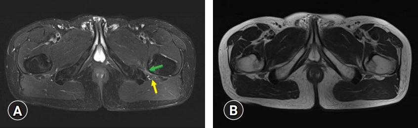
Fig. 1. (A) Pelvic axial fat-saturated T2-weighted and (B) T2-weighted magnetic resonance imaging (MRI) reveal that the ischiofemoral space is narrower on the left side than on the right side. Additionally, on the pelvic axial fat-saturated T2-weighted MRI (A), high signal intensity is found on the left quadratus femoris muscle (green arrow), indicative of muscle edema. Over the area of high signal intensity in the left quadratus femoris muscle, the left sciatic nerve is visible (yellow arrow).
Reference
-
References
1. Lee S, Kim I, Lee SM, Lee J. Ischiofemoral impingement syndrome. Ann Rehabil Med. 2013; 37:143–6.2. Ulusoy OL, Tutar S, Ozturk E, Mutlu A, Mutlu H. Ischiofemoral impingement syndrome: another cause of extraspinal sciatica. Spine J. 2016; 16:e527.3. Wilson MD, Keene JS. Treatment of ischiofemoral impingement: results of diagnostic injections and arthroscopic resection of the lesser trochanter. J Hip Preserv Surg. 2016; 3:146–53.
- Full Text Links
-
- Actions
-
Cited
- CITED
-
- Close
- Share
-
- Similar articles
-
- Atypical presentation of complex regional pain syndrome: neuropathic itching - A case report -
- Peripheral Effect of Morphine on Mechanical Allodynia in a Rat Model of Neuropathic Pain
- Effects of Cervical Sympathectomy on Mechanical Allodynia and Cold Allodynia in a Rat Model of Neuropathic Pain
- Clinical Scale for Neuropathic Pain
- Etiology and epidemiology of neuropathic pain
