Acute Crit Care.
2022 Aug;37(3):474-476. 10.4266/acc.2022.00339.
Contrast media mimicking subarachnoid hemorrhage after intrathecal injection in a patient with Creutzfeldt-Jakob disease
- Kim T
 1,2
1,2
- Affiliations
-
- 1Department of Emergency Medicine, Seoul National University Hospital, Seoul, Korea
- 2Department of Emergency Medicine, Seoul National University College of Medicine, Seoul, Korea
- KMID: 2535313
- DOI: http://doi.org/10.4266/acc.2022.00339
Figure
-
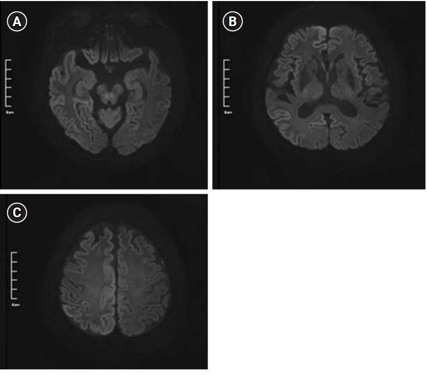
Figure 1. Brain diffusion-weighted imaging at b1000 at the levels of (A) midbrain, (B) thalamus, and (C) frontal and occipital lobes. Multifocal diffusion restriction can be seen along the bilateral cerebral cortex, especially prominent on the right parietal area.
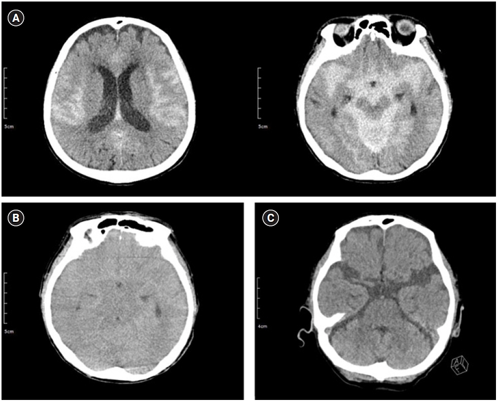
Figure 2. Serial brain computed tomography images. (A) At midnight on the day of fluoroscopy-guided cerebrospinal fluid drainage, diffuse high attenuation was seen in the subarachnoid space (66 HU). (B) Twelve hours after initial observation, diffuse sulcal high attenuation was still noted (59 HU); however, it was more dispersed into the subarachnoid space. (C) After 24 hours, high attenuation had nearly disappeared (21 HU). HU: hounsfield unit.
Reference
-
1. Hasan TF, Duarte W, Akinduro OO, Goldstein ED, Hurst R, Haranhalli N, et al. Nonaneurysmal "pseudo-subarachnoid hemorrhage" computed tomography patterns: challenges in an acute decision-making heuristics. J Stroke Cerebrovasc Dis. 2018; 27:2319–26.
Article2. Oh CH, An SD, Choi SH, Ji GY. Contrast mimicking a subarachnoid hemorrhage after lumbar percutaneous epidural neuroplasty: a case report. J Med Case Rep. 2013; 7:88.
Article3. Platt A, Ammar FE, Collins J, Ramos E, Goldenberg FD. Pseudo-subarachnoid hemorrhage and gadolinium encephalopathy following lumbar epidural steroid injection. Radiol Case Rep. 2020; 15:1935–8.
Article4. Sasaki Y, Ishii K, Ishii I. Myelography-associated pseudo-subarachnoid hemorrhage. Vis J Emerg Med. 2016; 5:25–6.
Article
- Full Text Links
-
- Actions
-
Cited
- CITED
-
- Close
- Share
-
- Similar articles
-
- Contrast media mimicking subarachnoid hemorrhage after intrathecal injection in a patient with Creutzfeldt-Jakob disease
- A case of Creutzfeldt-Jakob disease
- Creutzfeldt-Jakob Disease Mimicking a Stroke as Initial Manifestation
- New Variant Creutzfeldt-Jakob Disease
- Neurosyphilis Mimicking Limbic Encephalitis and Creutzfeldt-Jakob Disease
