PulseRider Treated Aneurysm with Significant Artifact on Postoperative Magnetic Resonance Angiography: A Case Report and Literature Review
- Affiliations
-
- 1Department of Neurosurgery, Baylor Scott & White Medical Center, Temple, TX, USA
- 2Department of Surgery, Texas A&M University College of Medicine, Temple, TX, USA
- KMID: 2522045
- DOI: http://doi.org/10.5469/neuroint.2021.00241
Abstract
- The PulseRider is a neuroendovascular adjunct for wide-necked intracranial aneurysms. The decreased metal burden of the PulseRider theoretically reduces artifact on radiologic imaging. However, we report here on a case of a patient who underwent PulseRider-assisted stent-coiling of a basilar tip aneurysm. He returned 19 months later for intermittent diplopia and darkening of vision but was neurologically intact on exam. Both contrast-enhanced and time-of-flight magnetic resonance angiography (MRA) demonstrated absence of signal in the basilar artery in the proximal anchors of the PulseRider. Given his lack of reproducible symptoms and high functional status, it is presumed that the imaging reflected artifact and not thrombosis/stenosis. Although the PulseRider is a useful treatment option for wide-necked intracranial aneurysms, the clinician should be aware that even contrast-enhanced MRA can produce artifact that resembles thrombosis/stenosis. Non-angiogram radiologic imaging modalities may be appropriate for evaluation for residual aneurysm but not patency of the parent artery.
Keyword
Figure
-
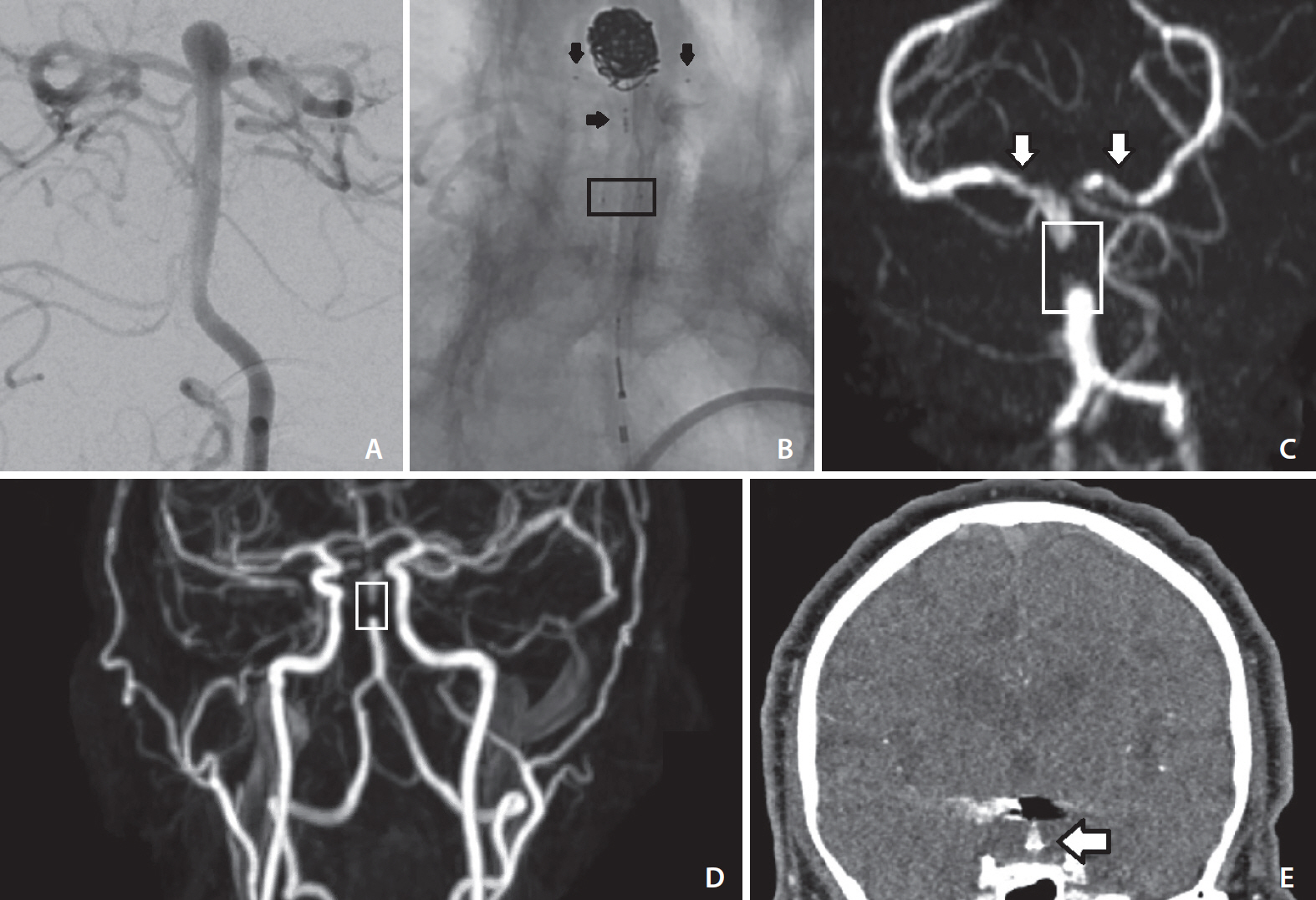
Fig. 1. Diagnostic cerebral angiogram demonstrating wide-necked basilar tip aneurysm (A). Diagnostic cerebral angiogram demonstrating patent basilar artery terminating in the bilateral posterior cerebral arteries status-post PulseRider-assisted coiling (B). Black horizontal arrows denote the medial markers of the PulseRider residing in the basilar artery, and black vertical arrows denote the markers of the PulseRider residing in the posterior cerebral arteries. A black box denotes the proximal radio-opaque markers of the PulseRider where the stainless-steel proximal anchors reside. Magnetic resonance angiography (MRA) of the head utilizing time-of-flight (TOF) techniques without contrast (C) and of the neck with contrast-enhanced techniques (D). In TOF-MRA, there is complete loss of signal at the proximal portion of the PulseRider (white box) and decreased but not absent signal within the proximal P1 segments of the bilateral posterior cerebral arteries (white arrows outlined in black). In CE-MRA, the artifact is again demonstrated in the proximal portion of the PulseRider (white box). There is signal distally without filling of the aneurysm. These results indicate that flow of the basilar artery to the posterior cerebral artery is not compromised and that there is no residual or recurrent aneurysm. A coronal reconstruction of computed tomographic angiography (CTA) demonstrates significant coil artifact and also two foci of PulseRider metal artifact resembling (white arrow outlined in black) in the proximal anchoring portion of the PulseRider (E). The stainless-steel retained in the proximal anchors are likely the culprit of the MRA and CTA artifact.
Reference
-
1. Hendricks BK, Yoon JS, Yaeger K, Kellner CP, Mocco J, De Leacy RA, et al. Wide-neck aneurysms: systematic review of the neurosurgical literature with a focus on definition and clinical implications. J Neurosurg. 2020; 133:159–165.
Article2. Spiotta AM, Chaudry MI, Turner RD 4th, Turk AS, Derdeyn CP, Mocco J, et al. An update on the adjunctive neurovascular support of wide-neck aneurysm embolization and reconstruction trial: 1-year safety and angiographic results. AJNR Am J Neuroradiol. 2018; 39:848–851.
Article3. Srinivasan VM, Srivatsan A, Spiotta AM, Hendricks BK, Ducruet AF, Albuquerque FC, et al. Early postmarket results with PulseRider for treatment of wide-necked intracranial aneurysms: a multicenter experience. J Neurosurg. 2020; 133:1756–1765.
Article4. Sakai N, Imamura H, Arimura K, Funatsu T, Beppu M, Suzuki K, et al. PulseRider-assisted coil embolization for treatment of intracranial bifurcation aneurysms: a single-center case series with 24-month follow-up. World Neurosurg. 2019; 128:e461–e467.
Article5. Gory B, Spiotta AM, Mangiafico S, Consoli A, Biondi A, Pomero E, et al. PulseRider stent-assisted coiling of wide-neck bifurcation aneurysms: periprocedural results in an international series. AJNR Am J Neuroradiol. 2016; 37:130–135.
Article6. Mukherjee S, Chandran A, Gopinathan A, Putharan M, Goddard T, Eldridge PR, et al. PulseRider-assisted treatment of widenecked intracranial bifurcation aneurysms: safety and feasibility study. J Neurosurg. 2017; 127:61–68.
Article7. Boddu SR, Tong FC, Dehkharghani S, Dion JE, Saindane AM. Contrast-enhanced time-resolved MRA for follow-up of intracranial aneurysms treated with the pipeline embolization device. AJNR Am J Neuroradiol. 2014; 35:2112–2118.
Article8. Anzalone N, Scomazzoni F, Cirillo M, Righi C, Simionato F, Cadioli M, et al. Follow-up of coiled cerebral aneurysms at 3T: comparison of 3D time-of-flight MR angiography and contrast-enhanced MR angiography. AJNR Am J Neuroradiol. 2008; 29:1530–1536.
Article9. Choi JW, Roh HG, Moon WJ, Kim NR, Moon SG, Kang CH, et al. Time-resolved 3D contrast-enhanced MRA on 3.0T: a non-invasive follow-up technique after stent-assisted coil embolization of the intracranial aneurysm. Korean J Radiol. 2011; 12:662–670.
Article10. Levent A, Yuce I, Eren S, Ozyigit O, Kantarci M. Contrast-enhanced and time-of-flight MR angiographic assessment of endovascular coiled intracranial aneurysms at 1.5 T. Interv Neuroradiol. 2014; 20:686–692.
Article11. De Leacy R, Yaniv G, Nael K. Cerebral aneurysm follow-up: how standards have changed and why. Endovasc Today. 2019; 18:80–84.12. Agid R, Schaaf M, Farb R. CE-MRA for follow-up of aneurysms post stent-assisted coiling. Interv Neuroradiol. 2012; 18:275–283.
Article13. Wang Y, Truong TN, Yen C, Bilecen D, Watts R, Trost DW, et al. Quantitative evaluation of susceptibility and shielding effects of nitinol, platinum, cobalt-alloy, and stainless steel stents. Magn Reson Med. 2003; 49:972–976.
Article
- Full Text Links
-
- Actions
-
Cited
- CITED
-
- Close
- Share
-
- Similar articles
-
- Artifacts in MR Angiography of the Intracranial Vessels Using the 3D TOF and 3D PC Techniques
- Magnetization Transfer Contrast Angiography for Organized Thrombosed Intracranial Aneurysm in TOF MR Angiography: a Case Report
- Ferromagnetic Artifact Due to Metallic Embolic Fragment after Endosaccular Coil Embolization: A Case Report
- Imaging Features of Intracranial Calcified Aneurysm: Report of 4 Cases
- Distal Middle Cerebral Artery M4 Aneurysm Surgery Using Navigation-CT Angiography
