Blood Res.
2020 Dec;55(4):275-278. 10.5045/br.2020.2020168.
Pulmonary embolism rate in patients infected with SARS-CoV-2
- Affiliations
-
- 1Division of Cardiothoracic Imaging, Department of Radiology, Columbia University Irving Medical Center, NY, USA
- 2Department of Biostatistics, Mailman School of Public Health, Columbia University, New York, NY, USA
- KMID: 2509966
- DOI: http://doi.org/10.5045/br.2020.2020168
Figure
-
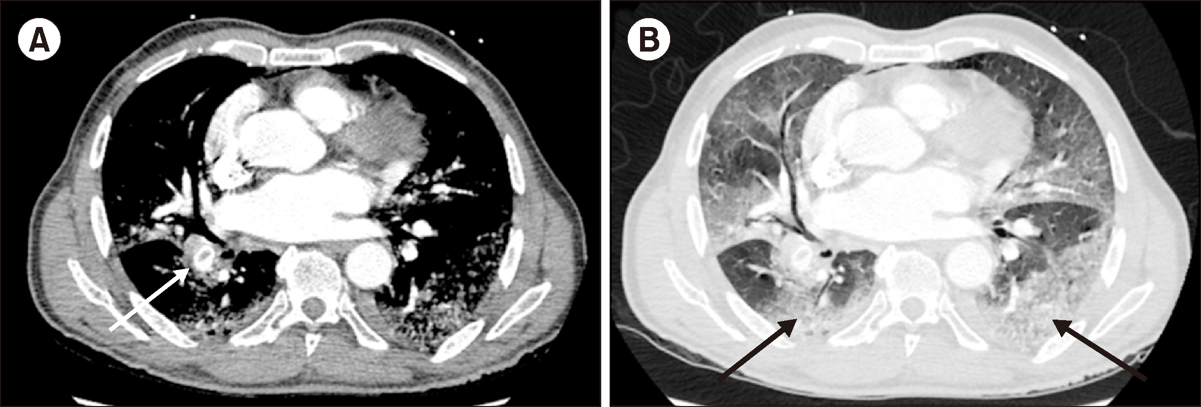
Fig. 1 This figure demonstrates an acute pulmonary embolism (white arrow) in a COVID-19 infected patient in soft tissue (A) and lung (B) windows. Pulmonary disease is characterized by peripheral and lower lobe predominant ground glass opacities (black arrows), typical of COVID-19 infection.
Reference
-
1. Chen N, Zhou M, Dong X, et al. 2020; Epidemiological and clinical characteristics of 99 cases of 2019 novel coronavirus pneumonia in Wuhan, China: a descriptive study. Lancet. 395:507–13. DOI: 10.1016/S0140-6736(20)30211-7. PMID: 32007143. PMCID: PMC7135076.
Article2. Klok FA, Kruip MJHA, van der Meer NJM, et al. 2020; Incidence of thrombotic complications in critically ill ICU patients with COVID-19. Thromb Res. 191:145–7. DOI: 10.1016/j.thromres.2020.04.013. PMID: 32291094. PMCID: PMC7146714.
Article3. Henrina J, Putra ICS, Cahyadi I, Gunawan HFH, Cahyadi A, Suciadi LP. 2020; Clinical characteristics and outcomes of venous thromboembolism in patients hospitalized for COVID-19: systematic review and meta-analysis. medRxiv. 20130922. DOI: 10.1101/2020.06.14.20130922.
Article4. Tian S, Hu W, Niu L, Liu H, Xu H, Xiao SY. 2020; Pulmonary pathology of early-phase 2019 novel coronavirus (COVID-19) pneumonia in two patients with lung cancer. J Thorac Oncol. 15:700–4. DOI: 10.1016/j.jtho.2020.02.010. PMID: 32114094. PMCID: PMC7128866.
Article5. Barton LM, Duval EJ, Stroberg E, Ghosh S, Mukhopadhyay S. 2020; COVID-19 autopsies, Oklahoma, USA. Am J Clin Pathol. 153:725–33. DOI: 10.1093/ajcp/aqaa062. PMID: 32275742. PMCID: PMC7184436.
Article6. Tian S, Xiong Y, Liu H, et al. 2020; Pathological study of the 2019 novel coronavirus disease (COVID-19) through postmortem core biopsies. Modern Pathol. 33:1007–14. DOI: 10.1038/s41379-020-0536-x. PMID: 32291399. PMCID: PMC7156231.
Article7. Dolhnikoff M, Duarte-Neto AN, de Almeida Monteiro RA, et al. 2020; Pathological evidence of pulmonary thrombotic phenomena in severe COVID-19. J Thromb Haemost. 18:1517–9. DOI: 10.1111/jth.14844. PMID: 32294295. PMCID: PMC7262093.
Article8. Schünemann HJ, Cushman M, Burnett AE, et al. 2018; American Society of Hematology 2018 guidelines for management of venous thromboembolism: prophylaxis for hospitalized and nonhospitalized medical patients. Blood Adv. 2:3198–225. DOI: 10.1182/bloodadvances.2018022954. PMID: 30482763. PMCID: PMC6258910.
Article9. Clerkin KJ, Fried JA, Raikhelkar J, et al. 2020; COVID-19 and cardiovascular disease. Circulation. 141:1648–55. DOI: 10.1161/CIRCULATIONAHA.120.046941. PMID: 32200663.
Article10. Allen J, Howell K. 2014; Microvascular imaging: techniques and opportunities for clinical physiological measurements. Physiol Meas. 35:R91–141. DOI: 10.1088/0967-3334/35/7/R91. PMID: 24910968.
Article11. Lang M, Som A, Mendoza DP, et al. 2020; Hypoxaemia related to COVID-19: vascular and perfusion abnormalities on dual-energy CT. Lancet Infect Dis. [Epub ahead of print]. DOI: 10.1016/S1473-3099(20)30367-4. PMID: 32359410. PMCID: PMC7252023.
Article
- Full Text Links
-
- Actions
-
Cited
- CITED
-
- Close
- Share
-
- Similar articles
-
- Intraoperative pulmonary embolism in shoulder arthroscopy in a patient with previous SARS-CoV-2 infection: a case report
- COVID-19 and Breastfeeding
- SARS-CoV-2 in the Prostate: Immunohistochemical and Ultrastructural Studies
- The Broad Spectrum of Neuro-Radiological Abnormalities in Patients Infected with SARS-CoV-2 Supports the Diagnosis of Neuro-COVID-19
- Understandings and Prospects of Laboratory Diagnosis of SARS-CoV-2
