Perianal Circumferential Reconstruction with Tailored Superior Gluteal Artery Perforator Flap
- Affiliations
-
- 1Department of Plastic Surgery, Samsung Medical Center, Seoul, Korea
- KMID: 2508945
- DOI: http://doi.org/10.12790/ahm.20.0032
Abstract
- Reconstruction following wide excision of perianal lesion is challenging as it requires resistance to high risks of wound contamination and preservation of anal function. Here, we present a case of a unilateral superior gluteal artery perforator (SGAP) flap with an opening in the flap. A 77-year old woman was referred due to an extramammary Pagets disease encircling the anus. Wide excision was performed by the general surgeon team, which generated a circumferential perianal defect. A unilateral SGAP flap was elevated. Primary defatting was done and an opening was made at the proper location of the anus. The anal mucosa was pulled out through the hole and sutured to the flap. She was discharged without any complications. At the follow-up visit, preservation of postoperative anal functions, as well as satisfactory contour, were observed. A well-tailored unilateral SGAP flap may be a good option for reconstruction of a perianal defect encircling the anus.
Figure
-
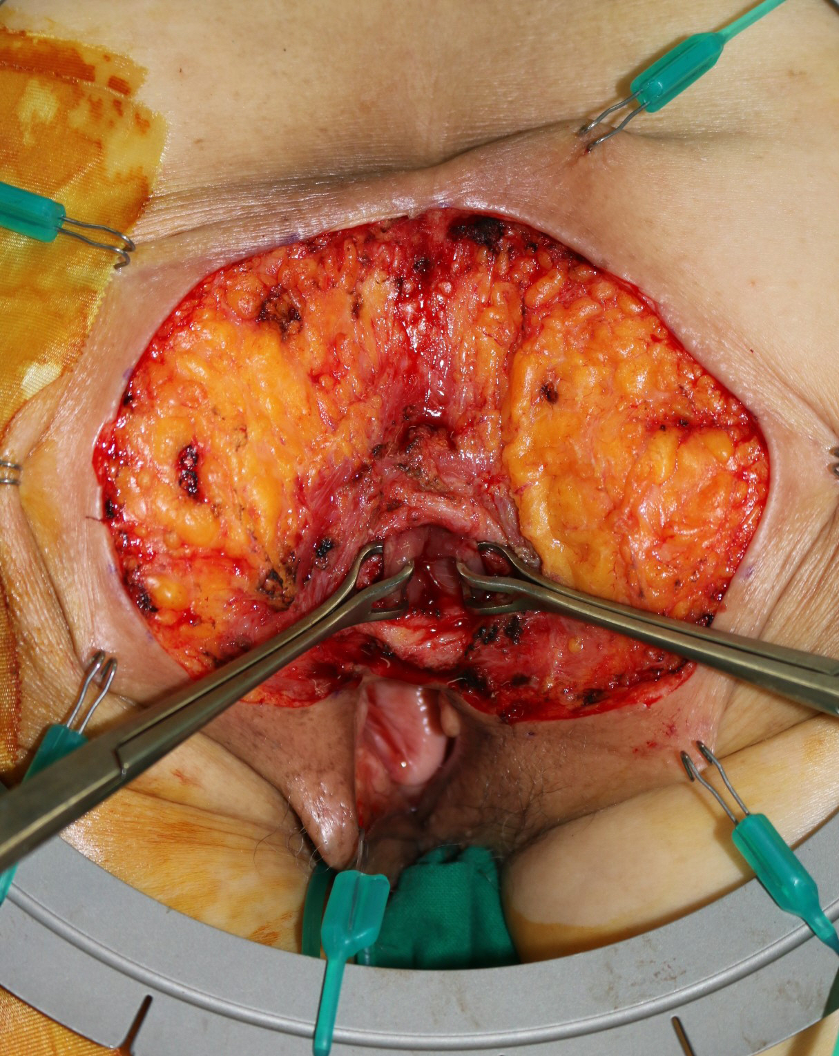
Fig. 1. Tissue defect encircling the anus, sized 10 × 5 cm. Written informed consent for publication of their clinical images was obtained from the patient.
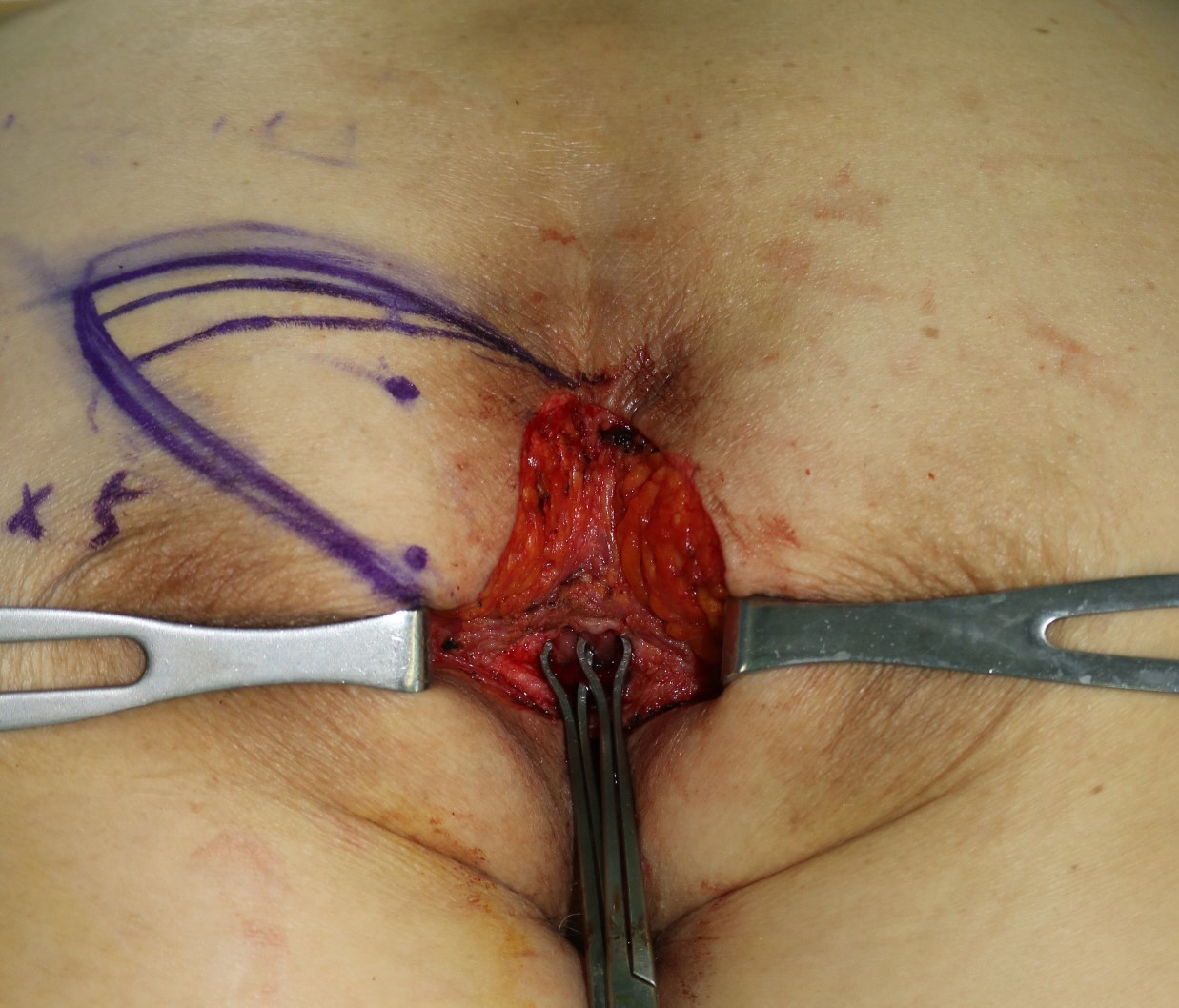
Fig. 2. Intraoperative design of the superior gluteal artery perforator flap from the left buttock based on three perforators. Written informed consent for publication of their clinical images was obtained from the patient.
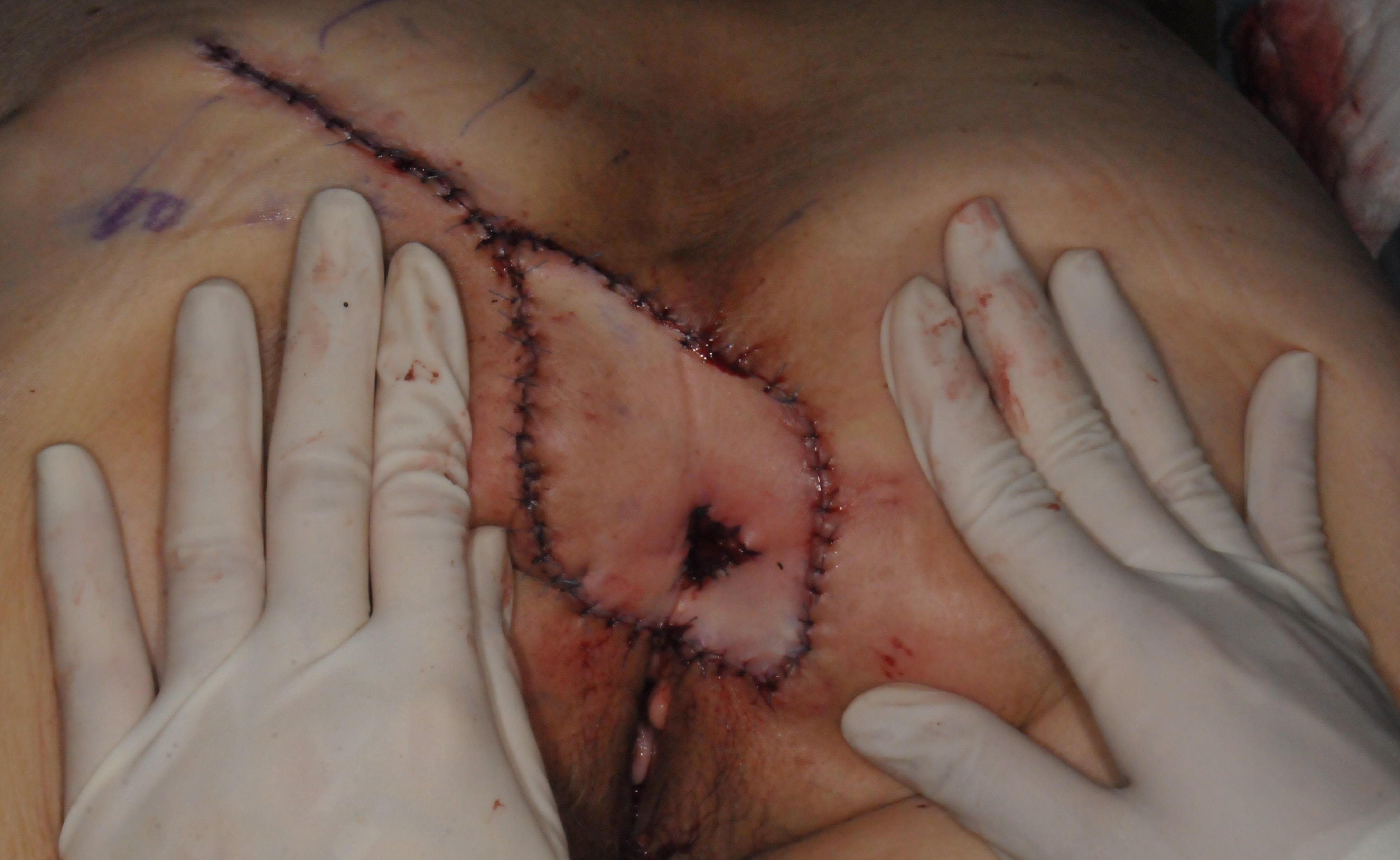
Fig. 3. Immediate postoperative photograph showing the anal mucosa sutured to the hole of the flap. Written informed consent for publication of their clinical images was obtained from the patient.

Fig. 4. Appearance at postoperative 3 months. Preservation of anal function and satisfactory tissue contouring is observed. Written informed consent for publication of their clinical images was obtained from the patient.
Reference
-
1. Ahmadzadeh R, Bergeron L, Tang M, Morris SF. The superior and inferior gluteal artery perforator flaps. Plast Reconstr Surg. 2007; 120:1551–6.
Article2. Borsuk DJ, Melich G, Sugrue J, et al. Wide local excision of perianal Paget’s disease with gluteal flap reconstruction: an interdisciplinary approach. J Vis Surg. 2016; 2:159.
Article3. Goto H, Yoshida Y, Kiyohara Y, Suyama Y, Nakayama B, Yamamoto O. Stoma creation for treatment of primary perianal Paget’s disease. Eur J Dermatol. 2015; 25:73–4.
Article4. Unal C, Yirmibesoglu OA, Ozdemir J, Hasdemir M. Superior and inferior gluteal artery perforator flaps in reconstruction of gluteal and perianal/perineal hidradenitis suppurativa lesions. Microsurgery. 2011; 31:539–44.
Article5. Sasaki K, Nozaki M, Kikutchi Y, Yamaki T, Soejima K. Reconstruction of perianal skin defect using a V-Y advancement of bilateral gluteus maximus musculocutaneous flaps: reconstruction considering anal cleft and anal function. Br J Plast Surg. 1999; 52:471–5.
Article6. Perez DR, Trakarnsanga A, Shia J, et al. Management and outcome of perianal Paget’s disease: a 6-decade institutional experience. Dis Colon Rectum. 2014; 57:747–51.
- Full Text Links
-
- Actions
-
Cited
- CITED
-
- Close
- Share
-
- Similar articles
-
- Dual Perforator Flap for Reconstruction of Large Sacral Defects: Superior Gluteal Artery Perforator Super-Flap with Parasacral Perforator
- The Treatment of Trochanteric Pressure Sore Using Superior or Inferior Gluteal Artery Perforator Flap
- Reconstruction of the Soft Tissue Defect Using Thoracodorsal Artery Perforator Skin Flap
- Treatment of Romberg`s Disease Using Superior Gluteal Perforator-based Flap
- A Superior Ulnar Collateral Artery Perforator Flap for a Large Defect on the Posterior Upper Arm
