J Pathol Transl Med.
2019 Sep;53(5):327-331. 10.4132/jptm.2019.05.14.
Amoebic Encephalitis Caused by Balamuthia mandrillaris
- Affiliations
-
- 1Department of Pathology, Keimyung University School of Medicine, Daegu, Korea. smkim5@kmu.ac.kr
- KMID: 2459581
- DOI: http://doi.org/10.4132/jptm.2019.05.14
Abstract
- We present the case of a 71-year-old man who was diagnosed with amoebic encephalitis caused by Balamuthia mandrillaris. He had rheumatic arthritis for 30 years and had undergone continuous treatment with immunosuppressants. First, he complained of partial spasm from the left thigh to the left upper limb. Magnetic resonance imaging revealed multifocal enhancing nodules in the cortical and subcortical area of both cerebral hemispheres, which were suggestive of brain metastases. However, the patient developed fever with stuporous mentality and an open biopsy was performed immediately. Microscopically, numerous amoebic trophozoites, measuring 20 to 25 µm in size, with nuclei containing one to four nucleoli and some scattered cysts having a double-layered wall were noted in the background of hemorrhagic necrosis. Based on the microscopic findings, amoebic encephalitis caused by Balamuthia mandrillaris was diagnosed. The patient died on the 10th day after being admitted at the hospital. The diagnosis of amoebic encephalitis in the early stage is difficult for clinicians. Moreover, most cases undergo rapid deterioration, resulting in fatal consequences. In this report, we present the first case of B. mandrillaris amoebic encephalitis with fatal progression in a Korean patient.
MeSH Terms
Figure
-
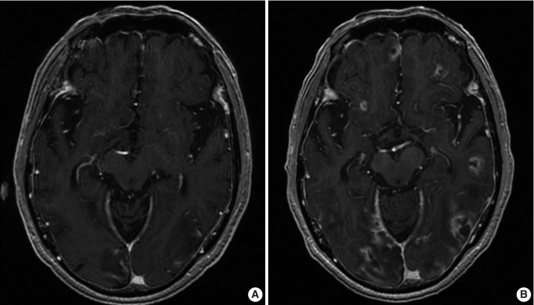
Fig. 1. (A) Initial magnetic resonance imaging (MRI) showing multiple ring-enhancing nodules in the cortical and subcortical areas of both cerebral hemispheres. (B) Second MRI showing an increased number and size of the nodules compared to the initial MRI.
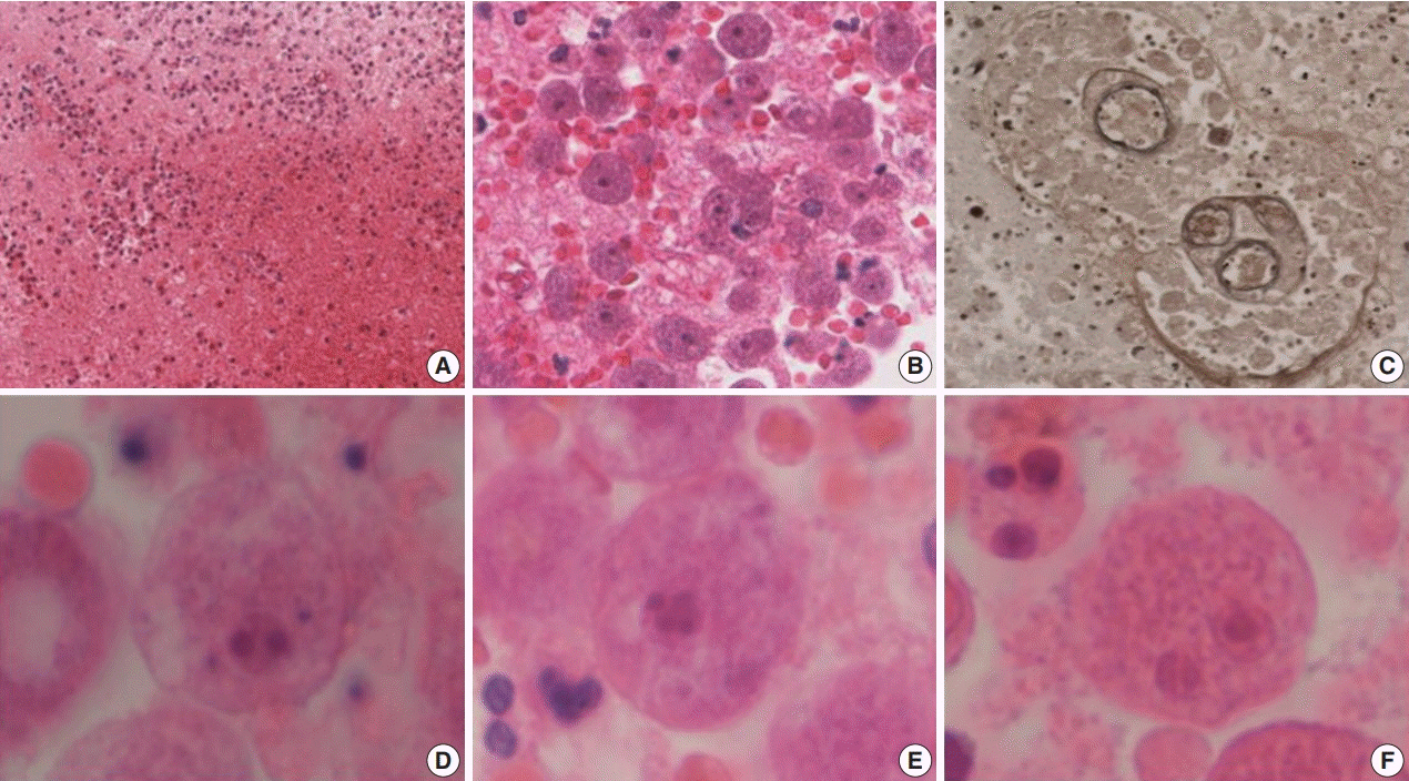
Fig. 2. Hemorrhagic necrosis (A) is associated with diffuse or perivascular infiltration of amoebic trophozoites (B, C). (C) Elastic stain highlights the trophozoites. Ovoid to round trophozoites, measuring 20 to 25 µm in size, with 1–2 nuclei containing 1–4 nucleoli are noted (D–F).
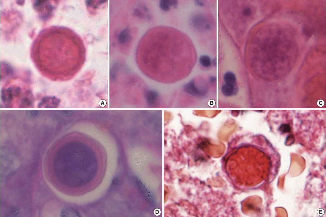
Fig. 3. (A–E) Spherical cysts, measuring 15–20 µm in size, consisting of a rigid double-layered wall are noted. The periodic-acid Schiff (D) and trichrome (E) stains highlight the cysts.
Reference
-
1. Martinez AJ. Infection of the central nervous system due to Acanthamoeba. Rev Infect Dis. 1991; 13 Suppl 5:S399–402.
Article2. Diaz JH. The public health threat from Balamuthia mandrillaris in the southern United States. J La State Med Soc. 2011; 163:197–204.3. Matin A, Siddiqui R, Jayasekera S, Khan NA. Increasing importance of Balamuthia mandrillaris. Clin Microbiol Rev. 2008; 21:435–48.4. Itoh K, Yagita K, Nozaki T, et al. An autopsy case of Balamuthia mandrillaris amoebic encephalitis, a rare emerging infectious disease, with a brief review of the cases reported in Japan. Neuropathology. 2015; 35:64–9.5. Diaz JH. Behavioral and recreational risk factors for free-living amebic infections. J Travel Med. 2011; 18:130–7.
Article6. Kiderlen AF, Laube U. Balamuthia mandrillaris, an opportunistic agent of granulomatous amebic encephalitis, infects the brain via the olfactory nerve pathway. Parasitol Res. 2004; 94:49–52.
Article7. Visvesvara GS, Schuster FL, Martinez AJ. Balamuthia mandrillaris, N. G., N. Sp., agent of amebic meningoencephalitis in humans and other animals. J Eukaryot Microbiol. 1993; 40:504–14.8. Visvesvara GS, Moura H, Schuster FL. Pathogenic and opportunistic free-living amoebae: Acanthamoeba spp., Balamuthia mandrillaris, Naegleria fowleri, and Sappinia diploidea. FEMS Immunol Med Microbiol. 2007; 50:1–26.9. Reddy R, Vijayasaradhi M, Uppin MS, Challa S, Jabeen A, Borghain R. Acanthamoeba meningoencephalitis in an immunocompetent patient: an autopsy case report. Neuropathology. 2011; 31:183–7.
- Full Text Links
-
- Actions
-
Cited
- CITED
-
- Close
- Share
-
- Similar articles
-
- Fatal Balamuthia Amebic Encephalitis in a Healthy Child: A Case Report with Review of Survival Cases
- Diagnosing Balamuthia mandrillaris amebic meningoencephalitis in a 64-year-old woman from the Southwest of China
- Fulminant Disseminating Fatal Granulomatous Amebic Encephalitis: The First Case Report in an Immunocompetent Patient in South Korea
- Generalized Peritonitis due to Acute Fulminant Amoebic Colitis
- Clinical Observation of Encephalitis Empasizing the Clinically Suspected Herpes Encephalitis Cases
