Single-Shot Echo-Planar Diffusion-Weighted MR Imaging at 3T and 1.5T for Differentiation of Benign Vertebral Fracture Edema and Tumor Infiltration
- Affiliations
-
- 1Department of Radiology, Kangbuk Samsung Hospital, Sungkyunkwan University School of Medicine, Seoul 03181, Korea. radiology11@daum.net
- KMID: 2458051
- DOI: http://doi.org/10.3348/kjr.2016.17.5.590
Abstract
OBJECTIVE
To compare the apparent diffusion coefficient (ADC) value using single-shot echo-planar imaging sequences at 3T and 1.5T for differentiation of benign fracture edema and tumor infiltration of the vertebral body.
MATERIALS AND METHODS
A total of 46 spinal examinations were included in the 1.5T MRI group, and a total of 40 spinal examinations were included in the 3T MRI group. The ADC values of the lesion were measured and calculated. The diagnostic performance of the conventional MR image containing sagittal T2-weighted fat saturated image and each diffusion weighted image (DWI) with an ADC value with different b values were evaluated.
RESULTS
The mean ADC value of the benign lesions was higher than that of the malignant lesions on 1.5T and 3T (p < 0.05). The sensitivity of the diagnostic performance was higher with an additional DWI in both 1.5T and 3T, but the sensitivities were similar with the addition of b values of 400 and 1000. The specificities of the diagnostic performances did not show significant differences (p value > 0.05). The diagnostic accuracies were higher when either of the DWIs (b values of 400 and 1000) was added to routine MR image for 1.5T and 3T. Statistical differences between 1.5T and 3T or between b values of 400 and 1000 were not seen.
CONCLUSION
The ADC values of the benign lesions were significantly higher than those of the malignant lesions on 1.5T and 3T. There was no statistically significant difference in the diagnostic performances when either of the DWIs (b values of 400 and 1000) was added to the routine MR image for 1.5T and 3T.
MeSH Terms
-
Adult
Aged
Aged, 80 and over
Bone Marrow Diseases/*diagnostic imaging
Diagnosis, Differential
Diffusion Magnetic Resonance Imaging/methods
Echo-Planar Imaging/methods
Edema/*diagnostic imaging
Female
Humans
Male
Middle Aged
Sensitivity and Specificity
Spinal Fractures/*diagnostic imaging
Spinal Neoplasms/diagnosis/*diagnostic imaging/secondary
Figure
-
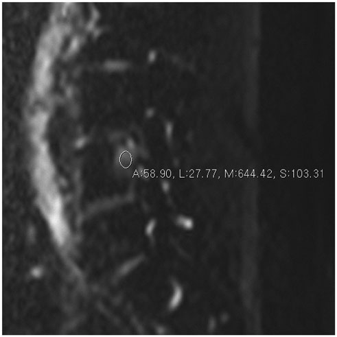
Fig. 1 Diffusion weighted image with b value of 0 in 60-year-old woman with lung cancer metastasis. Regions of interest were placed within lesion of vertebral body.
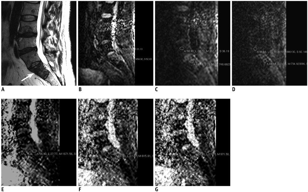
Fig. 2 58 years old man with oropharyngeal carcinoma metastasis. A. 3T MRI T2-weighted sagittal image (TR/TE, 2500/120) shows metastatic lesion in S1 (arrow). B. Diffusion weighted image with b value of 0. Regions of interest were placed within lesion (lower circle) and normal marrow (upper circle) of vertebral body. C. Diffusion weighted image with b value of 400. D. Diffusion weighted image with b value of 1000. E. ADC map with b value of 0–400. Region of interest were placed within lesion. F. ADC map with b value of 0–1000. G. ADC map with b value of 0–400–1000. First, both readers diagnosed lesion as malignant based on routine MR images. During second session, both readers diagnosed lesion as malignant based on routine MR images with additional information of DWI with b value 400. During third session, readers (both reader 1 and reader 2) diagnosed lesion as malignant based on routine MR images with additional information of DWI with b value 1000. ADC = apparent diffusion coefficient, DWI = diffusion weighted image, TE = echo time, TR = repetition time
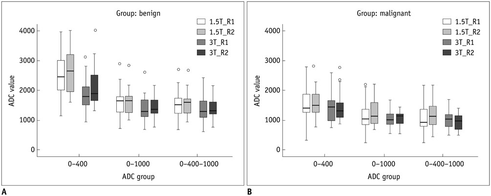
Fig. 3 Graph shows mean apparent diffusion coefficient (ADC) values (× 10-6 mm2/sec). A. Benign lesion. B. Malignant lesion. R1 = reader 1, R2 = reader 2
Cited by 2 articles
-
Diffusion-Weighted Magnetic Resonance Imaging of Spine
Young Cheol Yoon
J Korean Soc Radiol. 2020;81(1):58-69. doi: 10.3348/jksr.2020.81.1.58.MRI Evaluation of Suspected Pathologic Fracture at the Extremities from Metastasis: Diagnostic Value of Added Diffusion-Weighted Imaging
Sun-Young Park, Min Hee Lee, Ji Young Jeon, Hye Won Chung, Sang Hoon Lee, Myung Jin Shin
Korean J Radiol. 2019;20(5):812-822. doi: 10.3348/kjr.2018.0545.
Reference
-
1. Oztekin O, Ozan E, Hilal Adibelli Z, Unal G, Abali Y. SSH-EPI diffusion-weighted MR imaging of the spine with low b values: is it useful in differentiating malignant metastatic tumor infiltration from benign fracture edema? Skeletal Radiol. 2009; 38:651–658.2. Kim YP, Kannengiesser S, Paek MY, Kim S, Chung TS, Yoo YH, et al. Differentiation between focal malignant marrow-replacing lesions and benign red marrow deposition of the spine with T2*-corrected fat-signal fraction map using a three-echo volume interpolated breath-hold gradient echo Dixon sequence. Korean J Radiol. 2014; 15:781–791.3. Herneth AM, Philipp MO, Naude J, Funovics M, Beichel RR, Bammer R, et al. Vertebral metastases: assessment with apparent diffusion coefficient. Radiology. 2002; 225:889–894.4. Baur A, Stäbler A, Brüning R, Bartl R, Krödel A, Reiser M, et al. Diffusion-weighted MR imaging of bone marrow: differentiation of benign versus pathologic compression fractures. Radiology. 1998; 207:349–356.5. Castillo M. Diffusion-weighted imaging of the spine: is it reliable? AJNR Am J Neuroradiol. 2003; 24:1251–1253.6. Phalke VV, Gujar S, Quint DJ. Comparison of 3.0 T versus 1.5 T MR: imaging of the spine. Neuroimaging Clin N Am. 2006; 16:241–248. ix7. Ahlawat S, Khandheria P, Subhawong TK, Fayad LM. Differentiation of benign and malignant skeletal lesions with quantitative diffusion weighted MRI at 3T. Eur J Radiol. 2015; 84:1091–1097.8. Kuhl CK, Textor J, Gieseke J, von Falkenhausen M, Gernert S, Urbach H, et al. Acute and subacute ischemic stroke at high-field-strength (3.0-T) diffusion-weighted MR imaging: intraindividual comparative study. Radiology. 2005; 234:509–516.9. Rosner B. Fundamentals of biostatistics. 6th ed. Belmont, CA: Duxbury Press;2005.10. Viera AJ, Garrett JM. Understanding interobserver agreement: the kappa statistic. Fam Med. 2005; 37:360–363.11. Tang G, Liu Y, Li W, Yao J, Li B, Li P. Optimization of b value in diffusion-weighted MRI for the differential diagnosis of benign and malignant vertebral fractures. Skeletal Radiol. 2007; 36:1035–1041.12. Lang P, Wendland MF, Saeed M, Gindele A, Rosenau W, Mathur A, et al. Osteogenic sarcoma: noninvasive in vivo assessment of tumor necrosis with diffusion-weighted MR imaging. Radiology. 1998; 206:227–235.13. Spuentrup E, Buecker A, Adam G, van Vaals JJ, Guenther RW. Diffusion-weighted MR imaging for differentiation of benign fracture edema and tumor infiltration of the vertebral body. AJR Am J Roentgenol. 2001; 176:351–358.14. Castillo M, Arbelaez A, Smith JK, Fisher LL. Diffusion-weighted MR imaging offers no advantage over routine noncontrast MR imaging in the detection of vertebral metastases. AJNR Am J Neuroradiol. 2000; 21:948–953.15. Balliu E, Vilanova JC, Peláez I, Puig J, Remollo S, Barceló C, et al. Diagnostic value of apparent diffusion coefficients to differentiate benign from malignant vertebral bone marrow lesions. Eur J Radiol. 2009; 69:560–566.16. Geith T, Schmidt G, Biffar A, Dietrich O, Dürr HR, Reiser M, et al. Comparison of qualitative and quantitative evaluation of diffusion-weighted MRI and chemical-shift imaging in the differentiation of benign and malignant vertebral body fractures. AJR Am J Roentgenol. 2012; 199:1083–1092.17. Dietrich O, Biffar A, Reiser MF, Baur-Melnyk A. Diffusion-weighted imaging of bone marrow. Semin Musculoskelet Radiol. 2009; 13:134–144.18. Lee SY, Jee WH, Jung JY, Park MY, Kim SK, Jung CK, et al. Differentiation of malignant from benign soft tissue tumours: use of additive qualitative and quantitative diffusion-weighted MR imaging to standard MR imaging at 3.0 T. Eur Radiol. 2016; 26:743–754.19. Subhawong TK, Jacobs MA, Fayad LM. Diffusion-weighted MR imaging for characterizing musculoskeletal lesions. Radiographics. 2014; 34:1163–1177.20. Yuan YH, Xiao EH, He Z, Xiang J, Tang KL, Yan RH, et al. MR diffusion-weighed imaging of rabbit liver. World J Gastroenterol. 2005; 11:5506–5511.
- Full Text Links
-
- Actions
-
Cited
- CITED
-
- Close
- Share
-
- Similar articles
-
- Usefulness of Apparent Diffusion Coefficient in Ovarian Cystic Tumors Using Diffusion-Weighted Magnetic Resonance Imaging
- Analysis of Apparent Diffusion Coefficients of the Brain in Healthy Controls: A Comparison Study between Single-Shot Echo-Planar Imaging and Read-out-Segmented Echo-Planar Imaging
- Multi-slice Multi-echo Pulsed-gradient Spin-echo (MePGSE) Sequence for Diffusion Tensor Imaging MRI: A Preliminary Result
- Diagnostic Performance of Diffusion-Weighted Steady-State Free Precession in Differential Diagnosis of Neoplastic and Benign Osteoporotic Vertebral Compression Fractures: Comparison to Diffusion-Weighted Echo-Planar Imaging
- In Vivo and In Vitro Studies of the Steady State Free Precession-Diffusion-Weighted MR Imagings on Low b-value: Validation and Application to Bone Marrow Pathology
