Metastatic Neuroendocrine Tumor with Cardiac Involvement Utilizing Multi-Modality Imaging
- Affiliations
-
- 1Division of Cardiovascular Medicine, Mayo Clinic, Scottsdale, AZ, USA. Arsanjani.Reza@mayo.edu
- 2Division of Hematology/Medical Oncology, Mayo Clinic, Phoenix, AZ, USA.
- 3Department of Radiology, Mayo Clinic, Scottsdale, AZ, USA.
- KMID: 2454053
- DOI: http://doi.org/10.4070/kcj.2018.0367
Abstract
- No abstract available.
MeSH Terms
Figure
-
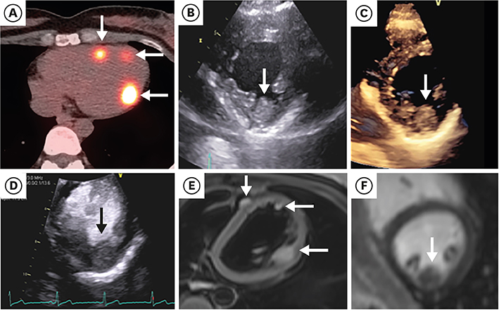
Figure 1 Myocardial metastasis of neuroendocrine tumor. (A) A Gallium-68 DOTATATE PET/CT hybrid image demonstrating radiotracer uptake in the heart (arrows). (B) Transthoracic images demonstrating large echogenic mass (arrow) along the inferolateral wall of the LV. (C) Three-dimensional image confirming presence of large mass (arrow). (D) Contrast-enhanced echocardiographic image demonstrating minimal contrast uptake indicating minimal vascularity (arrow). (E) Turbo spin T2 sequence demonstrating multiple high T2 signal along the LV and RV wall correlating well with Gallium-68 DOTATATE PET images (arrows). (F) There is minimal late gadolinium enhancement on the cardiac MRI (arrow). CT = computed tomography; LV = left ventricle; MRI = magnetic resonance imaging; PET = positron emission tomography; RV = right ventricular.
Reference
-
1. Davar J, Connolly HM, Caplin ME, et al. Diagnosing and managing carcinoid heart disease in patients with neuroendocrine tumors: an expert statement. J Am Coll Cardiol. 2017; 69:1288–1304.2. Bhattacharyya S, Toumpanakis C, Burke M, Taylor AM, Caplin ME, Davar J. Features of carcinoid heart disease identified by 2- and 3-dimensional echocardiography and cardiac MRI. Circ Cardiovasc Imaging. 2010; 3:103–111.
Article
- Full Text Links
-
- Actions
-
Cited
- CITED
-
- Close
- Share
-
- Similar articles
-
- Multi Modality Imaging Features of Cardiac Myxoma
- Skin Metastasis of Neuroendocrine Carcinoma Arising in the Rectum
- Incidental Muscle Uptake of 177 Lu-DOTATATE in Peripheral Vascular Disease
- Hypertriglyceridemia Associated with Use of Sunitinib to Treat a Metastatic Pancreatic Neuroendocrine Tumor
- Pancreas Neuroendocrine Tumor and Its Mimics: Review of Cross-Sectional Imaging Findings for Differential Diagnosis
