Complication Following Ultrasound-Guided High-Intensity Focused Ultrasound for the Treatment of Uterine Adenomyosis: Case Report of CT Imaging Features
- Affiliations
-
- 1Department of Radiology and Research Institute of Radiology, University of Ulsan College of Medicine, Asan Medical Center, Seoul, Korea. gshong@amc.seoul.kr
- 2Department of Radiology, Cheju Halla General Hospital, Jeju, Korea.
- KMID: 2454039
- DOI: http://doi.org/10.3348/jksr.2019.80.3.579
Abstract
- High intensity focused ultrasound (HIFU) is a non-surgical and non-invasive treatment option in patients with uterine myoma and adenomyosis. As the use of HIFU increases in the clinical practice, it is important to be aware of imaging findings related to ultrasound (US)-guided HIFU ablation and its potential complications. However, there are few reports on the imaging findings regarding complications of US-guided HIFU ablation. Here, we report a case of acute complication after US-guided HIFU ablation, surgically confirmed as thermal injury with necrosis of skin, subcutaneous tissue, anterior abdominal wall muscles, peritoneum and uterus.
MeSH Terms
Figure
-
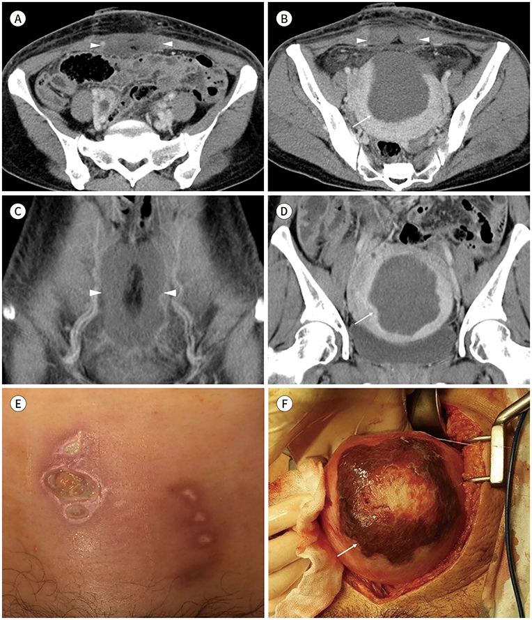
Fig. 1 Complication following ultrasound-guided HIFU ablation for uterine adenomyosis in a 44-year-old woman, presenting with low abdominal pain. A, B. Axial CT image shows overlying skin thickening, non-enhancing thickening of the superficial and deep fascia at the anterior pelvic wall, peritoneal thickening with fat infiltration in the pelvic cavity, and non-enhancement at bilateral rectus abdominis muscles (A, B; arrowheads). Note the sharply demarcated zone of hypodensity with linear lateral margins at the anterior portion of the uterus (B, arrow). C, D. Coronal reconstructed CT image shows area of non-enhancement at the bilateral rectus abdominis muscles (C, arrowheads) and zone of hypodensity at the anterior portion of the uterus (D, arrow). E, F. Photos show (E) skin necrosis in the treated area and (F) thermal injury with necrosis (arrow) on the uterine anterior wall in the surgical field. HIFU = high intensity focused ultrasound
Reference
-
1. Kim YS, Rhim H, Choi MJ, Lim HK, Choi D. High-intensity focused ultrasound therapy: an overview for radiologists. Korean J Radiol. 2008; 9:291–302.
Article2. Cheung VY. Current status of high-intensity focused ultrasound for the management of uterine adenomyosis. Ultrasonography. 2017; 36:95–102.
Article3. Hwang DW, Song HS, Kim HS, Chun KC, Koh JW, Kim YA. Delayed intestinal perforation and vertebral osteomyelitis after high-intensity focused ultrasound treatment for uterine leiomyoma. Obstet Gynecol Sci. 2017; 60:490–493.
Article4. Chen J, Chen W, Zhang L, Li K, Peng S, He M, et al. Safety of ultrasound-guided ultrasound ablation for uterine fibroids and adenomyosis: a review of 9988 cases. Ultrason Sonochem. 2015; 27:671–676.
Article5. Zhang L, Zhang W, Orsi F, Chen W, Wang Z. Ultrasound-guided high intensity focused ultrasound for the treatment of gynaecological diseases: a review of safety and efficacy. Int J Hyperthermia. 2015; 31:280–284.
Article6. Kim HK, Kim D, Lee MK, Lee CR, Kang SY, Chung YJ, et al. Three cases of complications after high-intensity focused ultrasound treatment in unmarried women. Obstet Gynecol Sci. 2015; 58:542–546.
Article7. Quinn SD, Vedelago J, Gedroyc W, Regan L. Safety and five-year re-intervention following magnetic resonance-guided focused ultrasound (MRgFUS) for uterine fibroids. Eur J Obstet Gynecol Reprod Biol. 2014; 182:247–251.
Article8. Ko JK, Seto MT, Cheung VY. Thermal bowel injury after ultrasound-guided high-intensity focused ultrasound for uterine adenomyosis. Ultrasound Obstet Gynecol. 2018; 52:282–283.9. Cheng CQ, Zhang , RT , Xiong Y, Chen L, Wang J, Huang GH, et al. Contrast-enhanced ultrasound for evaluation of high-intensity focused ultrasound treatment of benign uterine diseases: retrospective analysis of contrast safety. Medicine (Baltimore). 2015; 94:e729.10. Zhao WP, Chen JY, Chen WZ. Dynamic contrast-enhanced MRI serves as a predictor of HIFU treatment outcome for uterine fibroids with hyperintensity in T2-weighted images. Exp Ther Med. 2016; 11:328–334.
Article
- Full Text Links
-
- Actions
-
Cited
- CITED
-
- Close
- Share
-
- Similar articles
-
- Current status of high-intensity focused ultrasound for the management of uterine adenomyosis
- Spontaneous Uterine Rupture during Second Trimester Pregnancy after High-intensity Focused Ultrasound
- Efficacy, Efficiency, and Safety of Magnetic Resonance-Guided High-Intensity Focused Ultrasound for Ablation of Uterine Fibroids: Comparison with Ultrasound-Guided Method
- Expulsion of Fibroids to the Endometrial Cavity after Magnetic Resonance Imaging-guided High Intensity Focused Ultrasound Surgery (MRgFUS) Treatment of Intramural Uterine Fibroids
- Ultrasound imaging guided high intensity focused ultrasound (HIFU) may be a safe tool to ablate uterine myoma
