Anatomical Correlates of the "Closing-In" Phenomenon
- Affiliations
-
- 1Department of Neurology, Onnuri Hospital, Incheon, Korea.
- 2Graduate School of Medical Science and Engineering, KAIST, Daejeon, Korea.
- 3Department of Neurology, Asan Medical Center, University of Ulsan College of Medicine, Seoul, Korea.
- 4Department of Neurology, College of Medicine, The Catholic University of Korea, Seoul, Korea. ysshim@catholic.ac.kr, neuroman@catholic.ac.kr
- 5Department of Radiology, College of Medicine, The Catholic University of Korea, Seoul, Korea.
- 6Department of Neurology, Konyang University College of Medicine, Daejeon, Korea.
- KMID: 2443039
- DOI: http://doi.org/10.12779/dnd.2015.14.1.17
Abstract
- BACKGROUND AND PURPOSE
The "closing-in" phenomenon refers to the tendency to copy near or overlap a model while performing figure-copying tasks. The mechanisms underlying the closing-in phenomenon have not been fully elucidated, and previous studies only investigated the mechanisms through neuropsychological tests. We investigated the neuroanatomical correlates of the closing-in phenomenon using voxel-based morphometry (VBM).
METHODS
Thirty-eight patients diagnosed with probable Alzheimer's disease (AD) and 21 normal controls were included. All subjects underwent neuropsychological testing to diagnose dementia and magnetization prepared rapid acquisition gradient echo brain magnetic resonance imaging for the voxel-based statistical analysis. The subjects were asked to copy the modified Luria's alternating squares and triangles to quantify the closing-in phenomenon. We applied SPM8 for the VBM analysis to detect gray matter loss associated with the closing-in phenomenon.
RESULTS
The patients with probable AD showed a higher closing-in score than that of the normal control subjects (p<0.0001). The VBM analysis revealed more parietal and temporal atrophy in the patients with AD than that in the normal control group. Moreover, atrophy of the orbito-frontal area was associated with the closing-in phenomenon.
CONCLUSIONS
The closing-in phenomenon is dysfunction while performing figure-copying tasks and is more common in patients with AD. The analysis of the orbito-frontal area, which is associated with inhibiting primitive reflexes, revealed that the closing-in phenomenon is an imitation behavior commonly observed in patients with frontal lobe damage.
MeSH Terms
Figure
-

Fig. 1 Modified Luria's alternating squares and triangles.
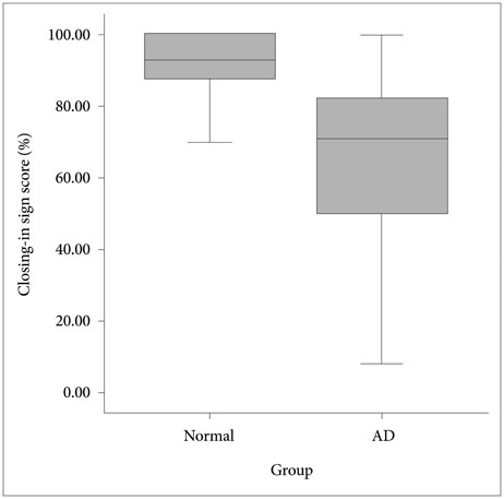
Fig. 2 Percent distribution of the closing-in scores for patients with Alzheimer's disease (AD) and the normal controls
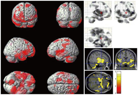
Fig. 3 Results of voxel-based morphometry t-test analysis comparing patterns of gray matter loss in the group of 38 patients with Alzheimer's disease (AD) with that of 21 healthy control subjects. Voxels showing significantly reduced gray matter volume in the patients with AD compared with the control group are indicated in red on a three-dimensional brain image (uncorrected p<0.05, left), in the gray scale on a glass-brain brain image (top right), and a template image with the color bar representing the t-statistic (corrected p<0.05, bottom right).
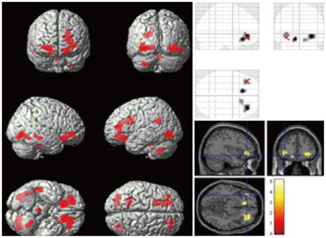
Fig. 4 Results of the voxel-based morphometry multiple regression analysis comparing patterns of gray matter loss associated with the closing-in phenomenon in 38 patients with Alzheimer's disease and 21 normal control subjects. Voxels show significantly reduced gray matter volume in the orbito-frontal areas in both groups (age and sex as covariates).
Reference
-
1. Gainotti G, Parlato V, Monteleone D, Carlomagno S. Neuropsychological markers of dementia on visual-spatial tasks: a comparison between Alzheimer's type and vascular forms of dementia. J Clin Exp Neuropsychol. 1992; 14:239–252.
Article2. Mayer-Gross W. Some Observations on Apraxia: (Section of Neurology). Proc R Soc Med. 1935; 28:1203–1212.3. Muncie W. Concrete model and abstract copy: a psychobiological interpretation of the "closing-in" symptom of Mayer-Gross. J Nerv Ment Dis. 1938; 88:1–11.4. de Ajuriaguerra, Muller M, Tissot R. [Apropos of some problems caused by apraxia in dementias]. Encephale. 1960; 49:375–401.5. Gainotti G. A quantitative study of the "closing-in" symptom in normal children and in brain-damaged patients. Neuropsychologia. 1972; 10:429–436.
Article6. Gainotti G, Marra C, Villa G, Parlato V, Chiarotti F. Sensitivity and specificity of some neuropsychological markers of Alzheimer dementia. Alzheimer Dis Assoc Disord. 1998; 12:152–162.
Article7. McIntosh RD, Ambron E, Della Sala S. Evidence for an attraction account of closing-in behaviour. Cogn Neuropsychol. 2008; 25:376–394.
Article8. Kwak YT. "Closing-in" phenomenon in Alzheimer's disease and subcortical vascular dementia. BMC Neurol. 2004; 4:3.
Article9. Lee BH, Chin J, Kang SJ, Kim EJ, Park KC, Na DL. Mechanism of the closing-in phenomenon in a figure copying task in Alzheimer's disease patients. Neurocase. 2004; 10:393–397.
Article10. Chin J, Lee BH, Seo SW, Kim EJ, Suh MK, Kang SJ, et al. The closing-in phenomenon in Alzheimer's disease and vascular dementia. J Clin Neurol. 2005; 1:166–173.
Article11. McKhann G, Drachman D, Folstein M, Katzman R, Price D, Stadlan EM. Clinical diagnosis of Alzheimer’s disease: report of the NINCDSADRDA Work Group under the auspices of Department of Health and Human Services Task Force on Alzheimer's Disease. Neurology. 1984; 34:939–944.
Article12. Choi SH, Na DL, Lee BH, Hahm DS, Jeong JH, Yoon SJ, et al. Estimating the validity of the Korean version of Expanded Clinical Dementia Rating (CDR) Scale. J Korean Neurol Assoc. 2001; 19:585–591.13. Kang Y, Na DL. Seoul Neuropsychological Screening Battery. Incheon: Human Brain Research & Consulting Co.;2003.14. Fazekas F, Chawluk JB, Alavi A, Hurtig HI, Zimmerman RA. MR signal abnormalities at 1.5 T in Alzheimer's dementia and normal aging. AJR Am J Roentgenol. 1987; 149:351–356.
Article15. Christensen KJ, Moye J, Armson RR, Kern TM. Health screening and random recruitment for cognitive aging research. Psychol Aging. 1992; 7:204–208.
Article16. Luria AR. Human brain and psychological processes. New York: Harper & Row;1966.17. Wright IC, McGuire PK, Poline JB, Travere JM, Murray RM, Frith CD, et al. A voxel-based method for the statistical analysis of gray and white matter density applied to schizophrenia. Neuroimage. 1995; 2:244–252.
Article18. Ashburner J, Friston KJ. Voxel-based morphometry--the methods. Neuroimage. 2000; 11(6 Pt 1):805–821.
Article19. Busatto GF, Diniz BS, Zanetti MV. Voxel-based morphometry in Alzheimer's disease. Expert Rev Neurother. 2008; 8:1691–1702.
Article20. Yang DW, Hong YJ, Yoon BR, Shim YS, Kim YI. Clinical use of Voxel-based morphometry. In : Monthly meeting of the Korean Dementia Association; Seoul. 2009. 2.21. Hänggi J, Streffer J, Jäncke L, Hock C. Volumes of lateral temporal and parietal structures distinguish between healthy aging, mild cognitive impairment, and Alzheimer’s disease. J Alzheimers Dis. 2011; 26:719–734.
Article22. Ambron E, Allaria F, McIntosh RD, Della Sala S. Closing-in behaviour in fronto-temporal dementia. J Neurol. 2009; 256:1004–1006.
Article23. Serra L, Fadda L, Perri R, Caltagirone C, Carlesimo GA. The closing-in phenomenon in the drawing performance of Alzheimer's disease patients: a compensation account. Cortex. 2010; 46:1031–1036.
Article24. Conson M, Salzano S, Manzo V, Grossi D, Trojano L. Closing-in without severe drawing disorders: the "fatal" consequences of pathological attraction. Cortex. 2009; 45:285–292.
Article25. Trojano L, Grossi D, Flash T. Cognitive neuroscience of drawing: contributions of neuropsychological, experimental and neurofunctional studies. Cortex. 2009; 45:269–277.
Article
- Full Text Links
-
- Actions
-
Cited
- CITED
-
- Close
- Share
-
- Similar articles
-
- Clinical Implication of "Closing-in" in Patients with Dementia
- The Closing-in Phenomenon in Alzheimer's Disease and Vascular Dementia
- Epileptiform Discharges on Closing the Eyes
- Detorque force and surface change of coated abutment screw after repeated closing and opening
- Severe Genu Recurvatum after a Closing-wedge High Tibial Osteotomy: A Case Report
