Chonnam Med J.
2019 Jan;55(1):62-63. 10.4068/cmj.2019.55.1.62.
Spinal Extradural Meningeal Cyst: A Case Report
- Affiliations
-
- 1Department of Neurosurgery, Chonnam National University Hospital, Chonnam National University Medical School, Gwangju, Korea. soohan@jnu.ac.kr
- KMID: 2432257
- DOI: http://doi.org/10.4068/cmj.2019.55.1.62
Abstract
- No abstract available.
Figure
-
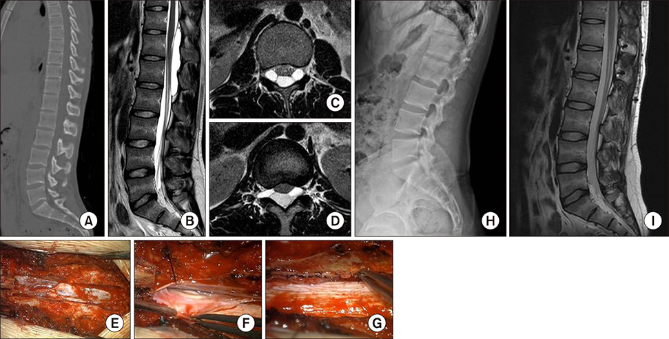
FIG. 1 Lumbar spine CT (A), sagittal T2-weighted MRI (B), axial T2-weighted MRI (C, D). Intraoperative photographs. After laminectomy (E), defect at dural sleeve near right T12 rootlet (F), primary closure of dural defect (G). 6-month follow-up after osteoplastic laminotomy and cyst removal. Plain lumbar radiography (H), sagittal T2-weighted MRI (I).
Reference
-
1. Chang IC. Surgical experience in symptomatic congenital intraspinal cysts. Pediatr Neurosurg. 2004; 40:165–170.
Article2. Lim MS, Khalil A, Okafo U, Dunlea O, Kaar G. Hemilaminectomy for large spinal extradural meningeal cysts: a case report and review of surgical techniques. Ann R Coll Surg Engl. 2016; 98:e162–e164.
Article3. Sangala JR, Uribe JS, Park P, Martinez C, Vale FL. Nerve root prolapse into a spinal arachnoid cyst--an unusual cause of radiculopathy. Clin Neurol Neurosurg. 2009; 111:460–464.
Article
- Full Text Links
-
- Actions
-
Cited
- CITED
-
- Close
- Share
-
- Similar articles
-
- The Spinal Extradural Meningeal Cyst Associated with High Disc Lumbar Herniation: Case Report
- Spinal Extradural Meningeal Cyst: Case Report
- A Case of The Surgically Treated Intraspinal Extradural Meningeal Cyst Demonstrating 'Ball-Valve' Mechanism of Formation
- Spinal Extradural Meningeal Cyst in Klippel-Trenaunay Syndrome
- Sacral Spinal Meningeal Cyst(Perineurial Cyst): A Case Report
