Upregulation of CD47 in Regulatory T Cells in Atopic Dermatitis
- Affiliations
-
- 1Department of Dermatology and Cutaneous Biology Research Institute, Yonsei University College of Medicine, Seoul, Korea. kwanglee@yuhs.ac
- 2Brain Korea 21 PLUS Project for Medical Science, Yonsei University College of Medicine, Seoul, Korea.
- 3Department of Dermatology, Yanbian University Hospital, Yanji, Jilin, China.
- 4Biomedical Group, Korea Basic Science Institute, Cheongju, Korea.
- KMID: 2427162
- DOI: http://doi.org/10.3349/ymj.2016.57.6.1435
Abstract
- PURPOSE
Regulatory T (Treg) cells are key modulators in the immune system. Recent studies have shown that atopic dermatitis (AD) patients have higher numbers of Treg cells; however, little is known about the specific phenotype and function of Treg cells in AD.
MATERIALS AND METHODS
To identify differentially expressed proteins in peripheral induced Treg cells in AD and naturally derived Treg cells in normal controls, CD4âºCD25⺠Treg cells were isolated from thymus tissue of normal mice and the spleens of AD mice. Membrane proteins were extracted, and quantitative proteomics labeling with Tandem Mass Tags (TMT) was performed, followed by one-dimensional liquid chromatography/tandem mass spectrometry analysis.
RESULTS
Using TMT labeling, we identified 510 proteins, including 63 membrane proteins and 16 plasma membrane proteins. CD47 was one of the upregulated proteins in Treg cells in AD spleens. Although CD47 was expressed in all CD4⺠and CD8⺠T cells, a significantly higher expression of CD47 was observed in the Treg cells of AD mice and AD patients than in those of normal mice and healthy controls. Furthermore, Treg cells from the spleen showed a significantly higher expression of CD47 than those from the thymus.
CONCLUSION
We found that CD47 is highly expressed in the Treg cells of AD mice, particularly in the spleen. Based on our results, we propose that CD47(high) Treg cells are likely induced Treg cells and that upregulated CD47 in the Treg cells of AD patients may play a role in the increased population of Treg cells in AD.
MeSH Terms
Figure
-
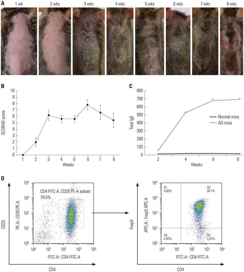
Fig. 1 Induction of AD-like skin lesions in NC/Nga mice and isolation of CD4+CD25+ Treg cells using MACS®. (A) Dorsal skin of mice treated with D. farinae extracts for 8 weeks. Significant erythema and crusts were observed after 3 weeks of D. farinae application. After 6 weeks of application, the most severe erythema, crusts, excoriation, and oozing were apparent. (B) SCORAD scores were plotted against the time of repeated topical application of D. farinae. (C) Serum IgE levels were measured via ELISA after repeated topical application of D. farinae ointment. All results are representative or mean±SD from groups that contained five mice. (D) CD4+CD25+ Treg cells were sorted using an AutoMACS cell sorter. The purity of isolated cells was 79.5%, and 94.7% of the isolated CD4+CD25+ Treg cells expressed Foxp3. Results are representative of three independent experiments. AD, atopic dermatitis; Treg, regulatory T; D. farinae, Dermatophagoides farinae; IgE, immunoglobulin E; ELISA, enzyme-linked immunosorbent assay.

Fig. 2 Expression of CD47 in various T cell subtypes. (A) FACS analysis of expression of CD47 in T cells, CD4+ T cells, and CD8+ T cells. All CD4+ T cells and CD8+ T cells expressed CD47. (B) FACS analysis of CD47 expression in CD3+CD4+CD25+ T cells and CD3+CD4+CD25+Foxp3+ T cells. Most CD4+CD25+Foxp3+ Treg cells expressed CD47. Treg, regulatory T; AD, atopic dermatitis.
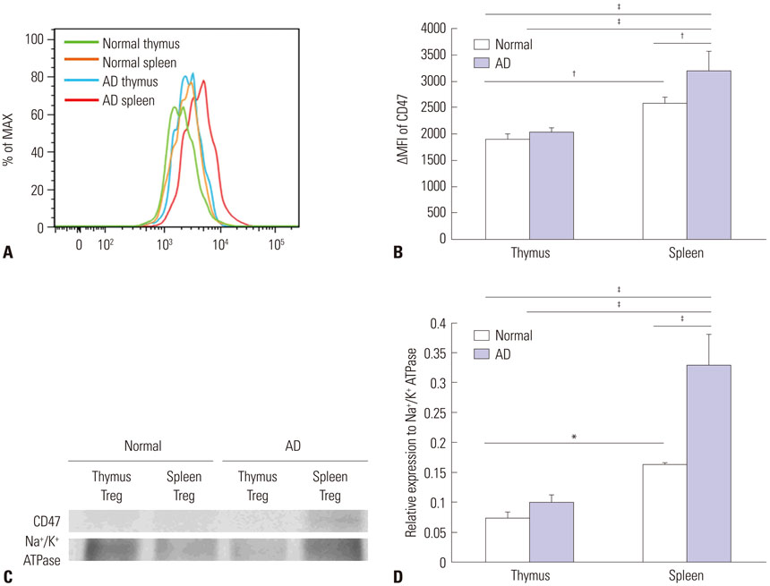
Fig. 3 Increased expression of CD47 in Treg cells from AD mice. (A) FACS analysis of expression of CD47 in Treg cells from thymuses and spleens from normal and AD mice. The ΔMFI of CD47 was determined in Treg cells. The green line indicates CD47 in a normal thymus, the orange line indicates CD47 in a normal spleen, the blue line indicates CD47 in an AD thymus, and the red line indicates CD47 in an AD spleen. (B) CD47 expression was more upregulated in Treg cells isolated from normal spleens than in those isolated from normal thymuses. Moreover, its expression was further upregulated in Treg cells from AD spleens. (C) Western blot analysis of the expression of CD47 on Treg cells from spleens and thymuses of AD mice and normal mice. The expression level of CD47 was similar to the results of the FACS analysis. (D) Western blot densitometry. Differences were determined via one-way ANOVA. For each group, mice n=4. *p<0.05, †p<0.01, ‡p<0.001. AD, atopic dermatitis; Treg, regulatory T.
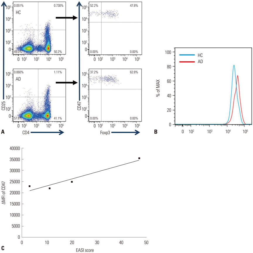
Fig. 4 Increased expression of CD47 in human Treg cells from AD patients. (A) All CD4+CD25+Foxp3+ Treg cells expressed CD47 in humans, a result similar to that for mice. (B) The expression level of CD47 was higher in Treg cells from AD patients than in those from healthy controls. (C) The severity of AD correlated with the expression of CD47 (Pearson correlation r=0.84, p=0.018). AD, atopic dermatitis; Treg, regulatory T.
Reference
-
1. Berke R, Singh A, Guralnick M. Atopic dermatitis: an overview. Am Fam Physician. 2012; 86:35–42.2. Furue M. Atopic dermatitis--immunological abnormality and its background. J Dermatol Sci. 1994; 7:159–168.
Article3. Guttman-Yassky E, Nograles KE, Krueger JG. Contrasting pathogenesis of atopic dermatitis and psoriasis--part I: clinical and pathologic concepts. J Allergy Clin Immunol. 2011; 127:1110–1118.
Article4. Guttman-Yassky E, Nograles KE, Krueger JG. Contrasting pathogenesis of atopic dermatitis and psoriasis--part II: immune cell subsets and therapeutic concepts. J Allergy Clin Immunol. 2011; 127:1420–1432.
Article5. Ohkura N, Kitagawa Y, Sakaguchi S. Development and maintenance of regulatory T cells. Immunity. 2013; 38:414–423.6. Ochs HD, Ziegler SF, Torgerson TR. FOXP3 acts as a rheostat of the immune response. Immunol Rev. 2005; 203:156–164.
Article7. Bacchetta R, Gambineri E, Roncarolo MG. Role of regulatory T cells and FOXP3 in human diseases. J Allergy Clin Immunol. 2007; 120:227–235.
Article8. Ito Y, Adachi Y, Makino T, Higashiyama H, Fuchizawa T, Shimizu T, et al. Expansion of FOXP3-positive CD4+CD25+ T cells associated with disease activity in atopic dermatitis. Ann Allergy Asthma Immunol. 2009; 103:160–165.
Article9. Gáspár K, Baráth S, Nagy G, Mócsai G, Gyimesi E, Szodoray P, et al. Regulatory T-cell subsets with acquired functional impairment: important indicators of disease severity in atopic dermatitis. Acta Derm Venereol. 2015; 95:151–155.
Article10. Samochocki Z, Alifier M, Bodera P, Jeziorkowska R, Rosiak E, Jurkiewicz B, et al. T-regulatory cells in severe atopic dermatitis: alterations related to cytokines and other lymphocyte subpopulations. Arch Dermatol Res. 2012; 304:795–801.
Article11. Szegedi A, Baráth S, Nagy G, Szodoray P, Gál M, Sipka S, et al. Regulatory T cells in atopic dermatitis: epidermal dendritic cell clusters may contribute to their local expansion. Br J Dermatol. 2009; 160:984–993.
Article12. Ou LS, Goleva E, Hall C, Leung DY. T regulatory cells in atopic dermatitis and subversion of their activity by superantigens. J Allergy Clin Immunol. 2004; 113:756–763.
Article13. Reefer AJ, Satinover SM, Solga MD, Lannigan JA, Nguyen JT, Wilson BB, et al. Analysis of CD25hiCD4+ "regulatory" T-cell subtypes in atopic dermatitis reveals a novel T(H)2-like population. J Allergy Clin Immunol. 2008; 121:415–422.14. Dayon L, Hainard A, Licker V, Turck N, Kuhn K, Hochstrasser DF, et al. Relative quantification of proteins in human cerebrospinal fluids by MS/MS using 6-plex isobaric tags. Anal Chem. 2008; 80:2921–2931.
Article15. Hanifin JM, Rajka G. Diagnostic features of atopic dermatitis. Acta Derm Venereol Suppl (Stockh). 1980; 92:44–47.16. Reinhold MI, Lindberg FP, Kersh GJ, Allen PM, Brown EJ. Costimulation of T cell activation by integrin-associated protein (CD47) is an adhesion-dependent, CD28-independent signaling pathway. J Exp Med. 1997; 185:1–11.
Article17. Han X, Sterling H, Chen Y, Saginario C, Brown EJ, Frazier WA, et al. CD47, a ligand for the macrophage fusion receptor, participates in macrophage multinucleation. J Biol Chem. 2000; 275:37984–37992.
Article18. Oldenborg PA, Zheleznyak A, Fang YF, Lagenaur CF, Gresham HD, Lindberg FP. Role of CD47 as a marker of self on red blood cells. Science. 2000; 288:2051–2054.
Article19. Brown EJ, Frazier WA. Integrin-associated protein (CD47) and its ligands. Trends Cell Biol. 2001; 11:130–135.
Article20. Gao AG, Lindberg FP, Finn MB, Blystone SD, Brown EJ, Frazier WA. Integrin-associated protein is a receptor for the C-terminal domain of thrombospondin. J Biol Chem. 1996; 271:21–24.
Article21. Lindberg FP, Bullard DC, Caver TE, Gresham HD, Beaudet AL, Brown EJ. Decreased resistance to bacterial infection and granulocyte defects in IAP-deficient mice. Science. 1996; 274:795–798.
Article22. Miyashita M, Ohnishi H, Okazawa H, Tomonaga H, Hayashi A, Fujimoto TT, et al. Promotion of neurite and filopodium formation by CD47: roles of integrins, Rac, and Cdc42. Mol Biol Cell. 2004; 15:3950–3963.
Article23. Jaiswal S, Jamieson CH, Pang WW, Park CY, Chao MP, Majeti R, et al. CD47 is upregulated on circulating hematopoietic stem cells and leukemia cells to avoid phagocytosis. Cell. 2009; 138:271–285.
Article24. Bouguermouh S, Van VQ, Martel J, Gautier P, Rubio M, Sarfati M. CD47 expression on T cell is a self-control negative regulator of type 1 immune response. J Immunol. 2008; 180:8073–8082.
Article25. Demeure CE, Tanaka H, Mateo V, Rubio M, Delespesse G, Sarfati M. CD47 engagement inhibits cytokine production and maturation of human dendritic cells. J Immunol. 2000; 164:2193–2199.
Article26. Doyen V, Rubio M, Braun D, Nakajima T, Abe J, Saito H, et al. Thrombospondin 1 is an autocrine negative regulator of human dendritic cell activation. J Exp Med. 2003; 198:1277–1283.
Article27. Latour S, Tanaka H, Demeure C, Mateo V, Rubio M, Brown EJ, et al. Bidirectional negative regulation of human T and dendritic cells by CD47 and its cognate receptor signal-regulator protein-alpha: down-regulation of IL-12 responsiveness and inhibition of dendritic cell activation. J Immunol. 2001; 167:2547–2554.
Article28. Avice MN, Rubio M, Sergerie M, Delespesse G, Sarfati M. CD47 ligation selectively inhibits the development of human naive T cells into Th1 effectors. J Immunol. 2000; 165:4624–4631.
Article29. Grimbert P, Bouguermouh S, Baba N, Nakajima T, Allakhverdi Z, Braun D, et al. Thrombospondin/CD47 interaction: a pathway to generate regulatory T cells from human CD4+ CD25- T cells in response to inflammation. J Immunol. 2006; 177:3534–3541.
Article30. Van VQ, Darwiche J, Raymond M, Lesage S, Bouguermouh S, Rubio M, et al. Cutting edge: CD47 controls the in vivo proliferation and homeostasis of peripheral CD4+ CD25+ Foxp3+ regulatory T cells that express CD103. J Immunol. 2008; 181:5204–5208.
Article31. Raimondi G, Turner MS, Thomson AW, Morel PA. Naturally occurring regulatory T cells: recent insights in health and disease. Crit Rev Immunol. 2007; 27:61–95.
Article32. Chen W, Jin W, Hardegen N, Lei KJ, Li L, Marinos N, et al. Conversion of peripheral CD4+CD25- naive T cells to CD4+CD25+ regulatory T cells by TGF-beta induction of transcription factor Foxp3. J Exp Med. 2003; 198:1875–1886.
Article33. Huber S, Stahl FR, Schrader J, Lüth S, Presser K, Carambia A, et al. Activin a promotes the TGF-beta-induced conversion of CD4+ CD25- T cells into Foxp3+ induced regulatory T cells. J Immunol. 2009; 182:4633–4640.
Article34. Bilate AM, Lafaille JJ. Induced CD4+Foxp3+ regulatory T cells in immune tolerance. Annu Rev Immunol. 2012; 30:733–758.35. Haribhai D, Lin W, Edwards B, Ziegelbauer J, Salzman NH, Carlson MR, et al. A central role for induced regulatory T cells in tolerance induction in experimental colitis. J Immunol. 2009; 182:3461–3468.
Article36. Oh SH, Park CO, Wu WH, Kim JY, Jin S, Byamba D, et al. Corticotropin-releasing hormone downregulates IL-10 production by adaptive forkhead box protein 3-negative regulatory T cells in patients with atopic dermatitis. J Allergy Clin Immunol. 2012; 129:151–159. 159.e1–159.e6.
Article37. Sugimoto N, Oida T, Hirota K, Nakamura K, Nomura T, Uchiyama T, et al. Foxp3-dependent and -independent molecules specific for CD25+CD4+ natural regulatory T cells revealed by DNA microarray analysis. Int Immunol. 2006; 18:1197–1209.
Article38. Hill JA, Feuerer M, Tash K, Haxhinasto S, Perez J, Melamed R, et al. Foxp3 transcription-factor-dependent and -independent regulation of the regulatory T cell transcriptional signature. Immunity. 2007; 27:786–800.
Article
- Full Text Links
-
- Actions
-
Cited
- CITED
-
- Close
- Share
-
- Similar articles
-
- A Study of the T cell Subset of Atopic Dermatitis
- Measurement of Atopic Dermatitis Disability
- Serum IgE Level in Patients of Atopic Dermatitis and Atopic Dermatitis with Molluscum Contagiosum
- Nipple Involvement in Atopic Dermatitis: Report of 3 cases
- Two Cases of Atopic Dermatitis Developing Ocular Complication and Immunological Disturbance
