Duodenal Amyloidosis
- Affiliations
-
- 1Department of Internal Medicine, Gangnam Severance Hospital, Yonsei University College of Medicine, Seoul, Korea. HJPARK21@yuhs.ac
- 2Department of Pathology, Gangnam Severance Hospital, Yonsei University College of Medicine, Seoul, Korea.
- KMID: 2416936
- DOI: http://doi.org/10.4166/kjg.2018.72.1.42
Abstract
- No abstract available.
MeSH Terms
Figure
-

Fig. 1. Esophagogastroduodenoscopic finding. (A) Numerous reddish, erosive lesions at duodenal bulb. (B-D) Numerous reddish, polypoid lesions at duodenal 2nd–3rd portion.
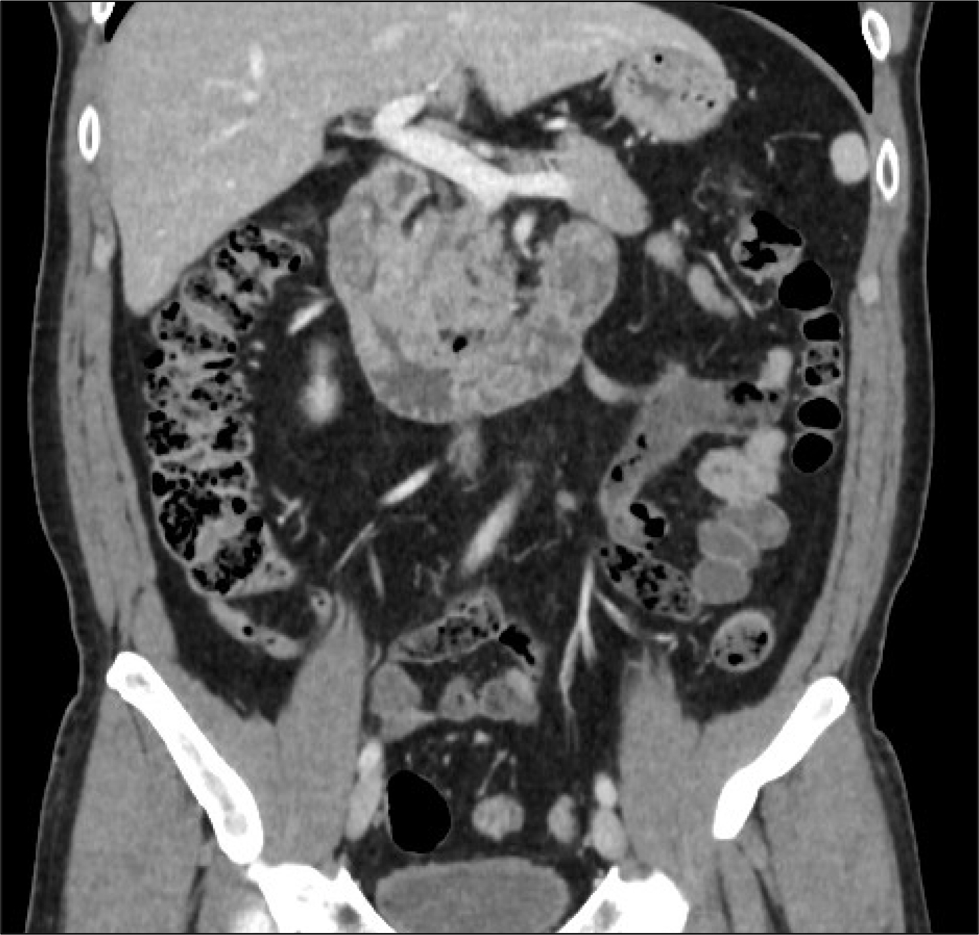
Fig. 2. Abdominal pevic CT. CT images show unusual fold thickening of the duodenal 2nd and 3rd portion. CT, computed tomography.

Fig. 3. (A, B) Histologic examination of duodenal mucosa. Amorphous pinkish material deposition in lamina propria of duodenal mucosa (A: hematoxylin and eosin stain [H&E], ×100; B: H&E, ×200). (C) Congo red stain shows the amorphous material with pale pink color (congo red stain, ×200) and (D) birefringence is not detected under polarized light (congo red stain, ×200).
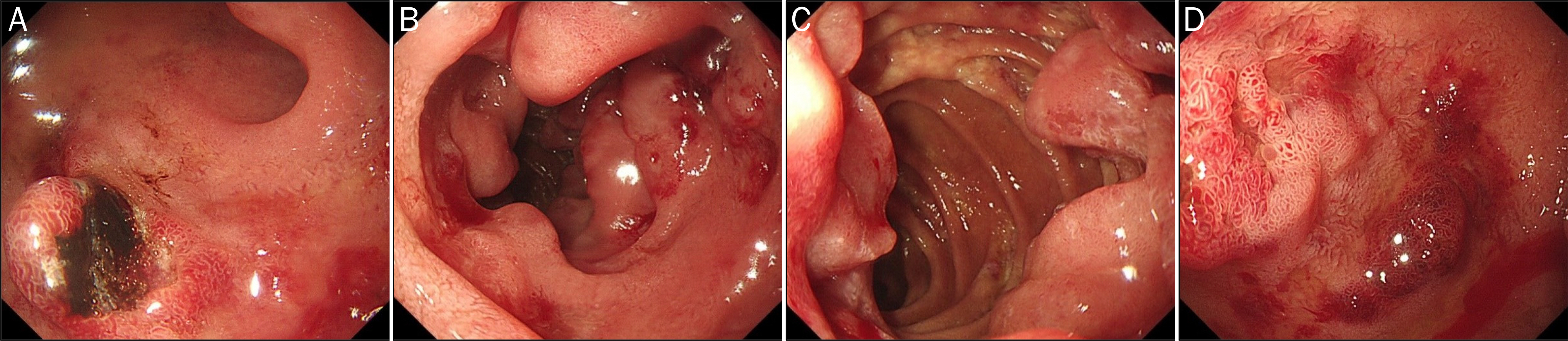
Fig. 4. Follow up esophagogastroduodenoscopic finding, 3 months later. (A) Polypoid and friable mucosa with hematin at duodenal bulb. (B-D) Numerous friable and polypoid mucosal lesion at duodenal 2nd–3rd portion.
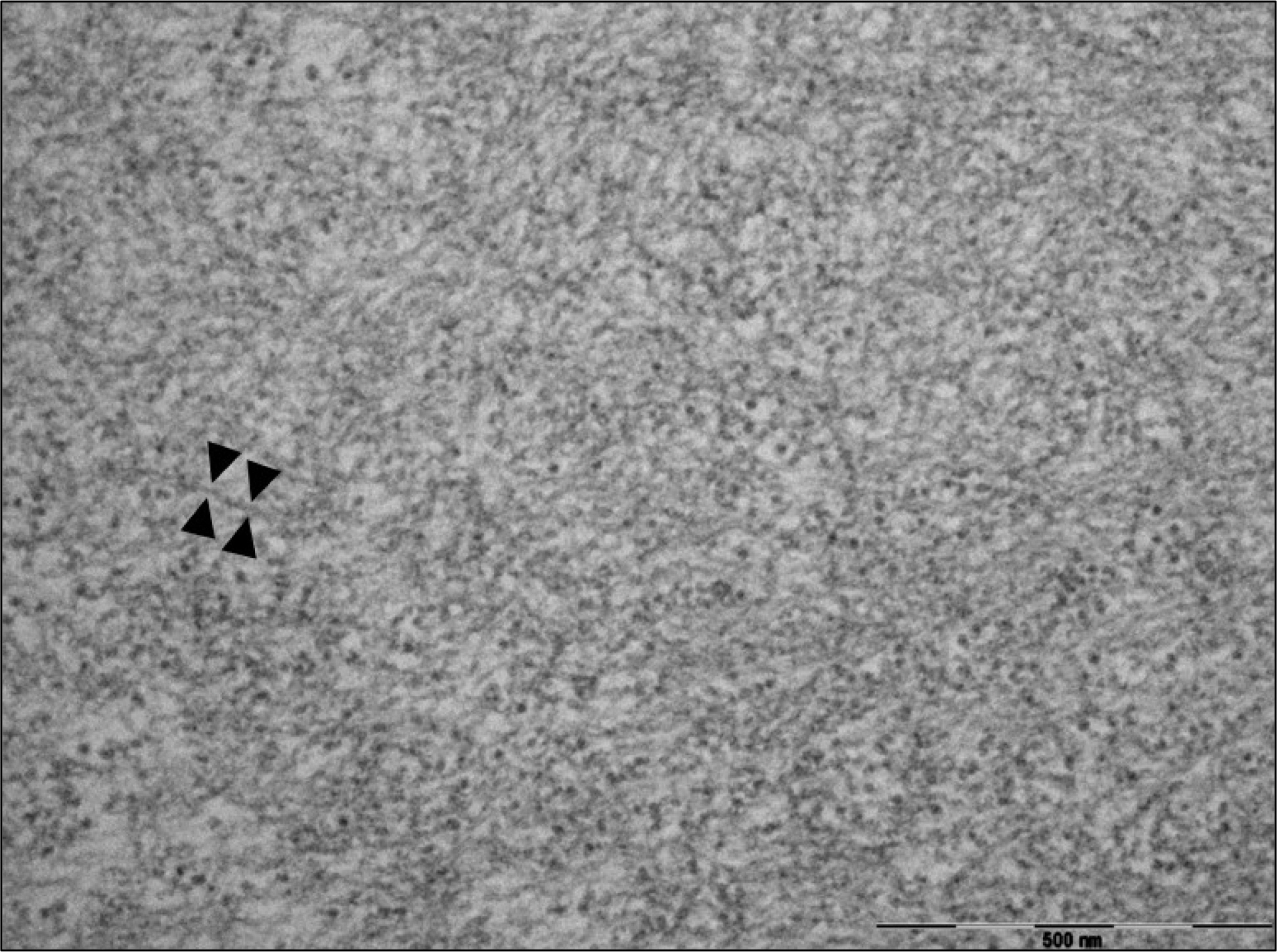
Fig. 5. High-power view of fibrils. The fibrils (between arrow heads) are randomly arranged in the loose background. The fibril size is ranged 7–13 nm (transmission electron microscopy ×80,000).
Reference
-
References
1. Kyle RA. Amyloidosis. Introduction and overview. J Intern Med. 1992; 232:507–508.2. Gaduputi V, Badipatla K, Patel H, Tariq H, Ihimoyan A. Primary systemic amyloidosis with extensive gastrointestinal involvement. Case Rep Gastroenterol. 2013; 7:511–515.
Article3. Sattianayagam PT, Hawkins PN, Gillmore JD. Systemic amyloidosis and the gastrointestinal tract. Nat Rev Gastroenterol Hepatol. 2009; 6:608–617.
Article4. Desport E, Bridoux F, Sirac C, et al. Al amyloidosis. Orphanet J Rare Dis. 2012; 7:54.
Article5. Gertz MA, Comenzo R, Falk RH, et al. Definition of organ involvement and treatment response in immunoglobulin light chain amyloidosis (AL): a consensus opinion from the 10th International Symposium on Amyloid and Amyloidosis, Tours, France, 18–22 April 2004. Am J Hematol. 2005; 79:319–328.6. Kim SY, Moon SB, Lee SK, et al. Light-chain amyloidosis presenting with rapidly progressive submucosal hemorrhage of the stomach. Asian J Surg. 2016; 39:113–115.
Article7. Cowan AJ, Skinner M, Seldin DC, et al. Amyloidosis of the gastrointestinal tract: a 13-year, single-center, referral experience. Haematologica. 2013; 98:141–146.
Article8. Hokama A, Kishimoto K, Nakamoto M, et al. Endoscopic and histopathological features of gastrointestinal amyloidosis. World J Gastrointest Endosc. 2011; 3:157–161.
Article
- Full Text Links
-
- Actions
-
Cited
- CITED
-
- Close
- Share
-
- Similar articles
-
- A Case of Duodenal Amyloidosis Accompanied with Candidiasis that was Diagnosed by Endoscopy
- AL amyloidosis: advances in diagnosis and management
- A Case of Nasopharyngeal Amyloidosis
- Primary Localized Amyloidosis of Bulbar Conjunctiva and Cornea
- Primary localized amyloidosis of the bladder: a case report
