Yeungnam Univ J Med.
2018 Jun;35(1):109-113. 10.12701/yujm.2018.35.1.109.
Coil embolization of ruptured intrahepatic pseudoaneurysm through percutaneous transhepatic biliary drainage
- Affiliations
-
- 1Department of Internal Medicine, College of Medicine, The Catholic University of Korea, Seoul, Korea.
- 2Department of Internal Medicine, Changwon Fatima Hospital, Changwon, Korea. 4796pjhsms@naver.com
- KMID: 2415743
- DOI: http://doi.org/10.12701/yujm.2018.35.1.109
Abstract
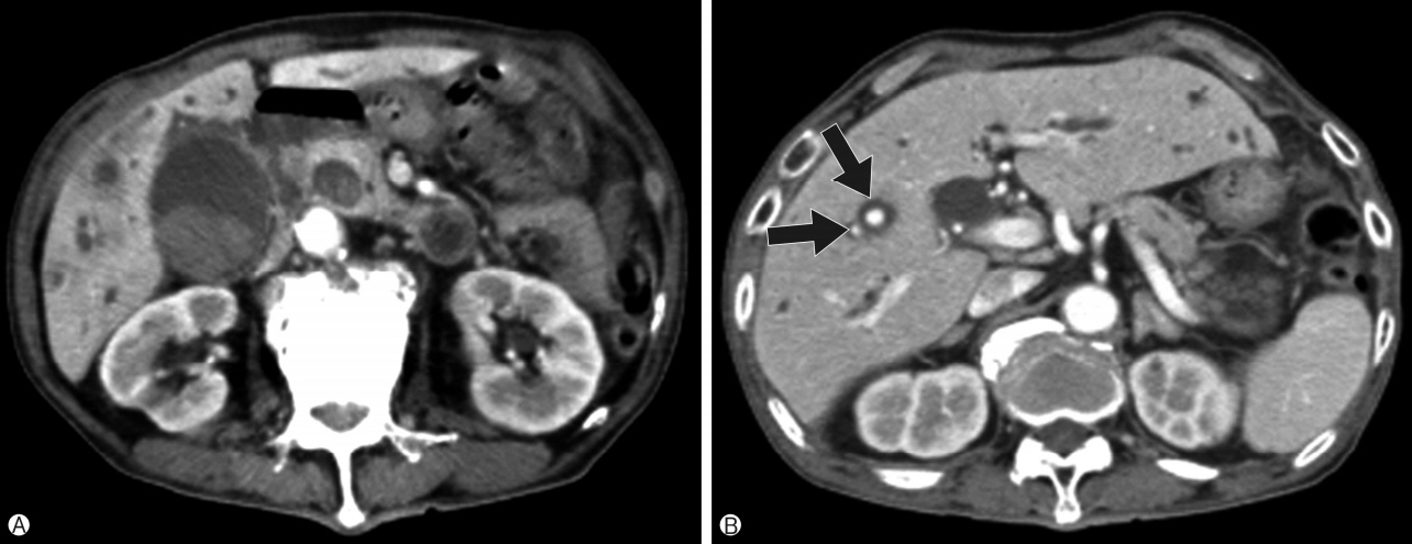
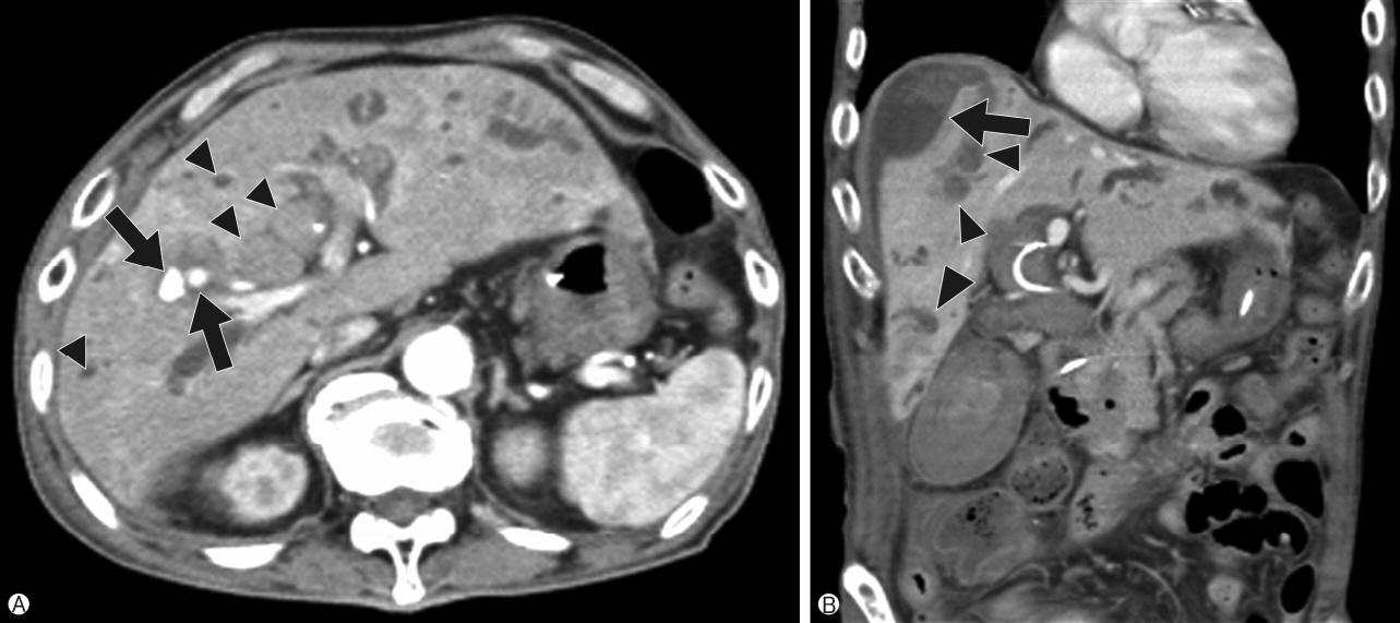
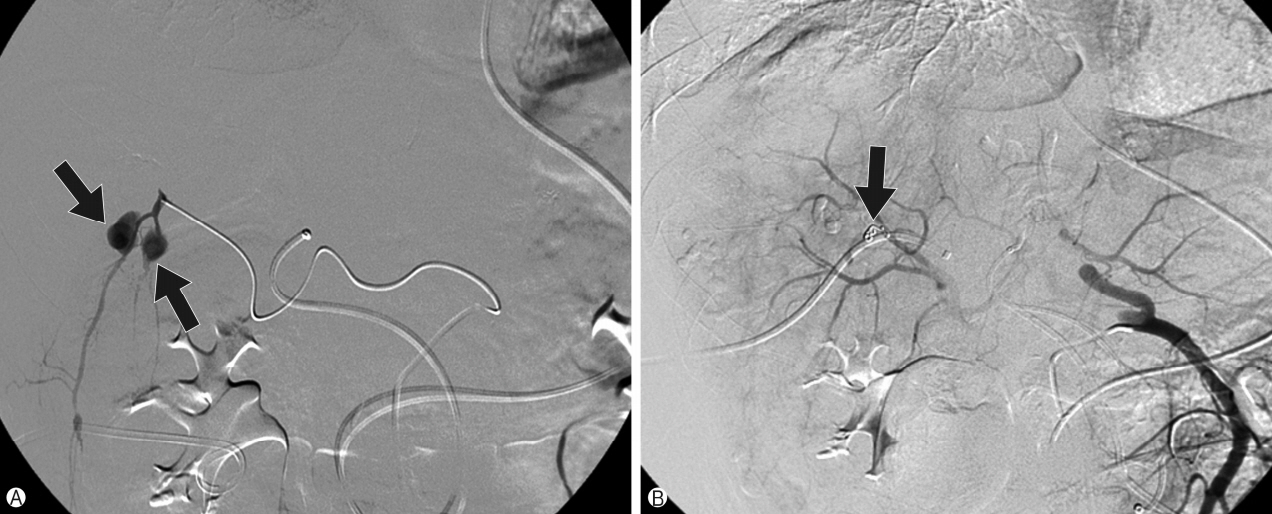
- A 75-year-old man with chronic cholangitis and a common bile duct stone that was not previously identified was admitted for right upper quadrant pain. Acute cholecystitis with cholangitis was suspected on abdominal computed tomography (CT); therefore, endoscopic retrograde cholangiopancreatography with endonasal biliary drainage was performed. On admission day 5, hemobilia with rupture of two intrahepatic artery pseudoaneurysms was observed on follow-up abdominal CT. Coil embolization of the pseudoaneurysms was conducted using percutaneous transhepatic biliary drainage. After several days, intrahepatic artery pseudoaneurysm rupture recurred and coil embolization through a percutaneous transhepatic biliary drainage tract was conducted after failure of embolization via the hepatic artery due to previous coiling. After the second coil embolization, a common bile duct stone was removed, and the patient presented no complications during 4 months of follow-up. We report a case of intrahepatic artery pseudoaneurysm rupture without prior history of intervention involving the hepatobiliary system that was successfully managed using coil embolization through percutaneous transhepatic biliary drainage.
Keyword
MeSH Terms
Figure
Reference
-
1. Nagaraja R, Govindasamy M, Varma V, Yadav A, Mehta N, Kumaran V, et al. Hepatic artery pseudoaneurysms: a singlecenter experience. Ann Vasc Surg. 2013; 27:743–9.
Article2. Willson TD, Korn JM, Blecha MJ, Podbielski FJ, Connolly MM. Spontaneous rupture of a saccular intrahepatic artery aneurysm. Vasc Endovascular Surg. 2012; 46:679–81.
Article3. Baggio E, Migliara B, Lipari G, Landoni L. Treatment of six hepatic artery aneurysms. Ann Vasc Surg. 2004; 18:93–9.
Article4. Dolapci M, Ersoz S, Kama NA. Hepatic artery aneurysm. Ann Vasc Surg. 2003; 17:214–6.
Article5. Yu YH, Sohn JH, Kim TY, Jeong JY, Han DS, Jeon YC, et al. Hepatic artery pseudoaneurysm caused by acute idiopathic pancreatitis. World J Gastroenterol. 2012; 18:2291–4.
Article6. Finley DS, Hinojosa MW, Paya M, Imagawa DK. Hepatic artery pseudoaneurysm: a report of seven cases and a review of the literature. Surg Today. 2005; 35:543–7.
Article7. Tessier DJ, Fowl RJ, Stone WM, McKusick MA, Abbas MA, Sarr MG, et al. Iatrogenic hepatic artery pseudoaneurysms: an uncommon complication after hepatic, biliary, and pancreatic procedures. Ann Vasc Surg. 2003; 17:663–9.
Article8. Lumsden AB, Mattar SG, Allen RC, Bacha EA. Hepatic artery aneurysms: the management of 22 patients. J Surg Res. 1996; 60:345–50.
Article9. Abbas MA, Fowl RJ, Stone WM, Panneton JM, Oldenburg WA, Bower TC, et al. Hepatic artery aneurysm: factors that predict complications. J Vasc Surg. 2003; 38:41–5.
Article10. Cho GH, Lee JJ, Yu SK, Kwon KA, Park DK, Kim YS, et al. A case of non-traumatic hemobilia due to pseudoaneurysm of the hepatic artery. Korean J Gastrointest Endosc. 2006; 33:173–7. Korean.11. Razik R, Fallah A, Sandroussi C, Wei AC, McGilvray ID. Dissecting pseudoaneurysm of the proper hepatic artery repaired by primary anastomosis: a case report. Case Rep Surg. 2012; 2012:804919.
Article
- Full Text Links
- Actions
-
Cited
- CITED
-
- Close
- Share
- Similar articles
-
- Percutaneous transhepatic biliary drainage
- Percutaneous transheptic removal of biliary stones:clinical analysis of 16 cases
- Percutaneous Transhepatic Electrohydraulic Lithotripsy for Stones in Billiary Tracts
- Percutaneous biliary drainage in acute suppurative cholangitis with biliary sepsis
- Percutaneous transhepatic biliary drainage


