Contrast Enhanced Harmonic Endoscopic Ultrasound: A Novel Approach for Diagnosis and Management of Gastrointestinal Stromal Tumors
- Affiliations
-
- 1Department of Internal Medicine, Yale-Waterbury Internal Medicine Program, Yale school of medicine, Waterbury, CT, USA.
- 2Division of Gastroenterology and Hepatology, Department of Digestive Diseases and Transplantation, Einstein Healthcare Network, Philadelphia, PA, USA.
- 3Department of Radiology, University of Texas Health Science Center at Houston, Houston, TX, USA.
- 4Division of Gastroenterology, Hepatology and Nutrition, University of Texas Health Science Center at Houston, Houston, TX, USA. sdsinghal@gmail.com
- KMID: 2414861
- DOI: http://doi.org/10.5946/ce.2017.170
Abstract
- The histologic analysis of gastrointestinal stromal tumors (GISTs) is a common method to detect the mitotic activity and to subsequently determine the risk of GISTs for malignancy. The potential false negative error due to inadequate yield of specimens and actual determination of malignancy risk requires analysis of the whole tumor. We aimed to assess the role of contrast enhanced endoscopic ultrasound (CE-EUS) in the management of GISTs. Two authors individually did review of English literatures to identify nine peer-reviewed original articles using keywords- contrast endoscopic ultrasound, GIST and submucosal tumor. Studies were heterogeneous in their aims looking either at differentiating submucosal lesions from GISTs, estimating malignant potential of GISTs with histologic correlation or studying the role of angiogenesis in malignant risk stratification. CE-EUS had moderate to high efficacy in differentiating GISTs from alternative submucosal tumors. CE-EUS had a higher sensitivity than EUS-guided fine needle aspiration, contrast computed tomography and Doppler EUS for detection of neo-vascularity within the GISTs. However, the evidence of abnormal angiogenesis within GIST as a prognostic factor needs further validation. CE-EUS is a non-invasive modality, which can help differentiate GISTs and provide valuable assessment of their perfusion patterns to allow better prediction of their malignant potential but more experience is needed.
MeSH Terms
Figure
-
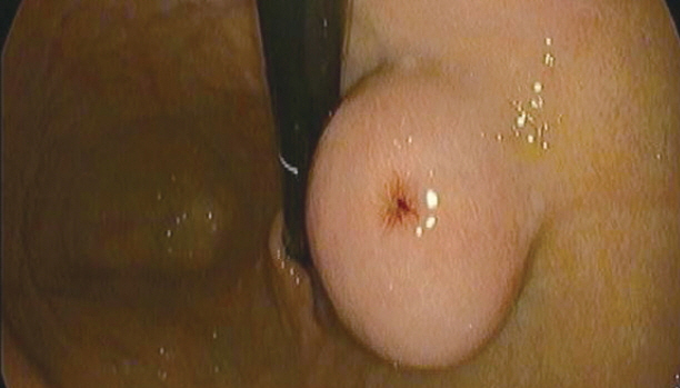
Fig. 1. Endoscopic view of submucosal tumor.
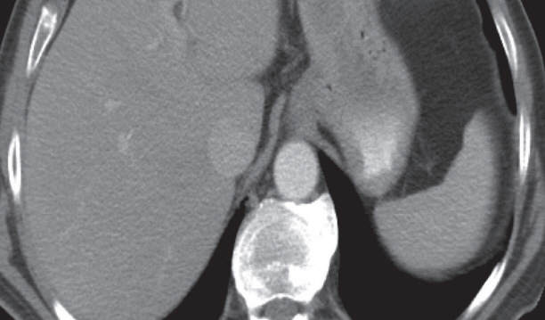
Fig. 2. Contrast enhanced computed tomography axial image showing a well-circumscribed submucosal lesion along the lesser curvature of the stomach without necrosis.
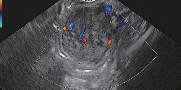
Fig. 3. Endoscopic ultrasound with Doppler showing vascular flow.
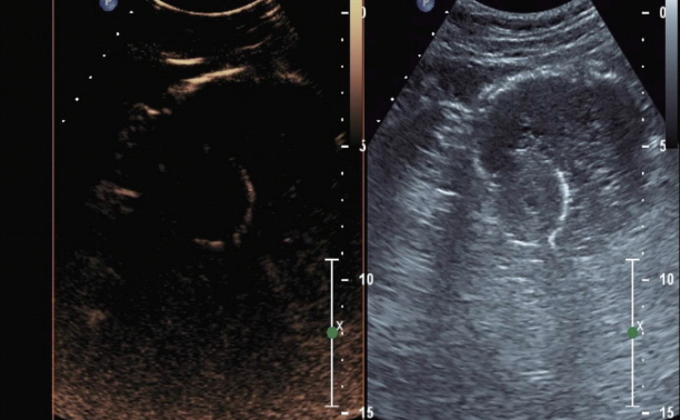
Fig. 4. Contrast enhanced harmonic endoscopic ultrasound dual image at the start of contrast administration- left image representing contrast image and the right image representing a tissue image.
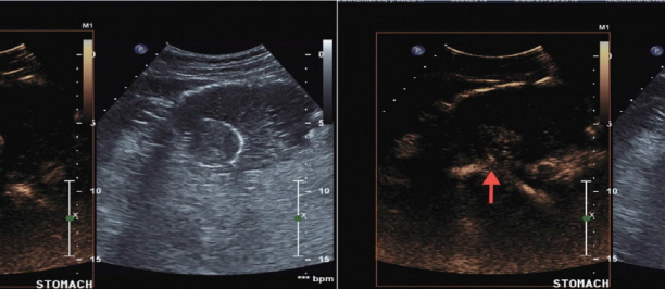
Fig. 5. Contrast enhanced harmonic endoscopic ultrasound images immediately after contrast administration- neo-vascularity (red arrow) in the submucosal lesion.
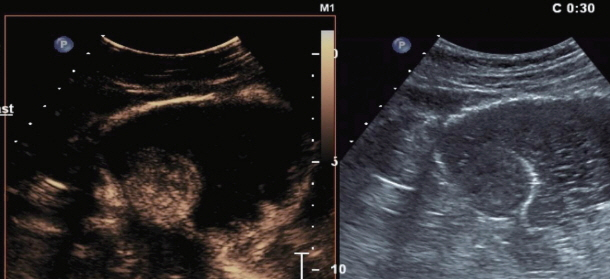
Fig. 6. Contrast enhanced harmonic endoscopic ultrasound images at 30 seconds after contrast administration- intense enhancement of the submucosal lesion.
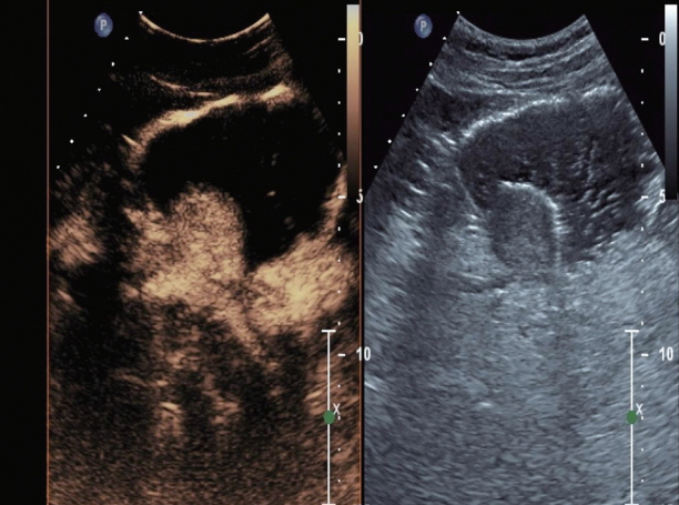
Fig. 7. Contrast enhanced harmonic endoscopic ultrasound images at 39 seconds after contrast administration- further enhancement of the submucosal lesion.
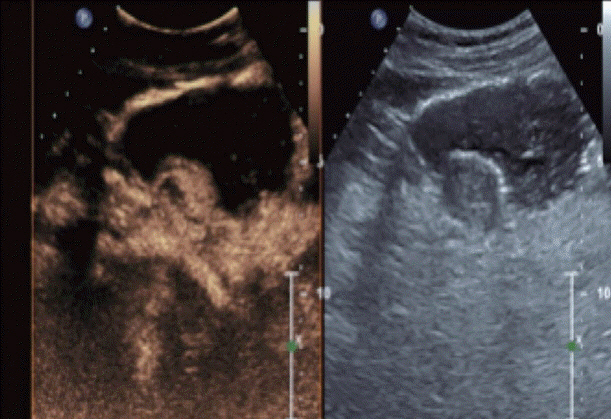
Fig. 8. Contrast enhanced harmonic endoscopic ultrasound images depicting focal area of necrosis within the submucosal lesion.
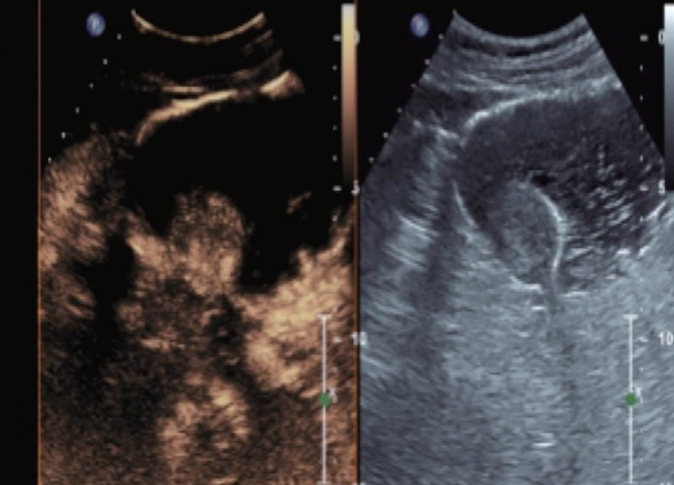
Fig. 9. Contrast enhanced harmonic endoscopic ultrasound images at 60 seconds after contrast administration- washout of the submucosal lesion.
Cited by 2 articles
-
Efficacy and Safety of Endoscopic Treatment for Gastrointestinal Stromal Tumors in the Upper Gastrointestinal Tract
Cicilia Marcella, Shakeel Sarwar, Hui Ye, Rui Hua Shi
Clin Endosc. 2020;53(4):458-465. doi: 10.5946/ce.2019.121.Contrast Enhanced Endoscopic Ultrasound Imaging for Gastrointestinal Subepithelial Tumors
Takashi Tamura, Masayuki Kitano
Clin Endosc. 2019;52(4):306-313. doi: 10.5946/ce.2019.056.
Reference
-
1. Kim KM, Kang DW, Moon WS, et al. Gastrointestinal stromal tumors in Koreans: it’s incidence and the clinical, pathologic and immunohistochemical findings. J Korean Med Sci. 2005; 20:977–984.
Article2. Goettsch WG, Bos SD, Breekveldt-Postma N, Casparie M, Herings RM, Hogendoorn PC. Incidence of gastrointestinal stromal tumours is underestimated: results of a nation-wide study. Eur J Cancer. 2005; 41:2868–2872.
Article3. Croom KF, Perry CM. Imatinib mesylate: in the treatment of gastrointestinal stromal tumours. Drugs. 2003; 63:513–522. discussion 523-524.4. Rammohan A, Sathyanesan J, Rajendran K, et al. A gist of gastrointestinal stromal tumors: a review. World J Gastrointest Oncol. 2013; 5:102–112.
Article5. Fletcher CD, Berman JJ, Corless C, et al. Diagnosis of gastrointestinal stromal tumors: a consensus approach. Int J Surg Pathol. 2002; 10:81–89.
Article6. Chak A, Canto MI, Rösch T, et al. Endosonographic differentiation of benign and malignant stromal cell tumors. Gastrointest Endosc. 1997; 45:468–473.
Article7. Kim GH, Kim KB, Lee SH, et al. Digital image analysis of endoscopic ultrasonography is helpful in diagnosing gastric mesenchymal tumors. BMC Gastroenterol. 2014; 14:7.
Article8. Kim GH, Park DY, Kim S, et al. Is it possible to differentiate gastric GISTs from gastric leiomyomas by EUS? World J Gastroenterol. 2009; 15:3376–3381.
Article9. Desser TS, Jeffrey RB. Tissue harmonic imaging techniques: physical principles and clinical applications. Semin Ultrasound CT MR. 2001; 22:1–10.
Article10. Kollmann C. New sonographic techniques for harmonic imaging--underlying physical principles. Eur J Radiol. 2007; 64:164–172.
Article11. Sanchez MV, Varadarajulu S, Napoleon B. EUS contrast agents: what is available, how do they work, and are they effective? Gastrointest Endosc. 2009; 69(2 Suppl):S71–S77.12. Săftoiu A, Dietrich CF, Vilmann P. Contrast-enhanced harmonic endoscopic ultrasound. Endoscopy. 2012; 44:612–617.
Article13. Wilson SR, Greenbaum LD, Goldberg BB. Contrast-enhanced ultrasound: what is the evidence and what are the obstacles? AJR Am J Roentgenol. 2009; 193:55–60.
Article14. Unnikrishnan S, Klibanov AL. Microbubbles as ultrasound contrast agents for molecular imaging: preparation and application. AJR Am J Roentgenol. 2012; 199:292–299.
Article15. Kitano M, Sakamoto H, Matsui U, et al. A novel perfusion imaging technique of the pancreas: contrast-enhanced harmonic EUS (with video). Gastrointest Endosc. 2008; 67:141–150.
Article16. Kitano M, Kudo M, Sakamoto H, et al. Preliminary study of contrast-enhanced harmonic endosonography with second-generation contrast agents. J Med Ultrason (2001). 2008; 35:11–18.
Article17. Zhao Y, Qian L, Li P, Zhang S. The diagnostic value of endoscopic ultrasonography and contrast-enhanced harmonic endoscopic ultrasonography in gastrointestinal stromal tumors. Endosc Ultrasound. 2016; 5:111–117.
Article18. Ignee A, Jenssen C, Hocke M, et al. Contrast-enhanced (endoscopic) ultrasound and endoscopic ultrasound elastography in gastrointestinal stromal tumors. Endosc Ultrasound. 2017; 6:55–60.
Article19. Kannengiesser K, Mahlke R, Petersen F, et al. Contrast-enhanced harmonic endoscopic ultrasound is able to discriminate benign submucosal lesions from gastrointestinal stromal tumors. Scand J Gastroenterol. 2012; 47:1515–1520.
Article20. Sakamoto H, Kitano M, Matsui S, et al. Estimation of malignant potential of GI stromal tumors by contrast-enhanced harmonic EUS (with videos). Gastrointest Endosc. 2011; 73:227–237.
Article21. Fukuta N, Kitano M, Maekawa K, Chikugo T, Kudo M. Estimation of the malignant potential of gastrointestinal stromal tumors: the value of contrast-enhanced coded phase-inversion harmonics US. J Gastroenterol. 2005; 40:247–255.
Article22. Yamashita Y, Kato J, Ueda K, et al. Contrast-enhanced endoscopic ultrasonography can predict a higher malignant potential of gastrointestinal stromal tumors by visualizing large newly formed vessels. J Clin Ultrasound. 2015; 43:89–97.
Article23. Park HY, Jeon SW, Lee HS, et al. Can contrast-enhanced harmonic endosonography predict malignancy risk in gastrointestinal subepithelial tumors? Endosc Ultrasound. 2016; 5:384–389.
Article
- Full Text Links
-
- Actions
-
Cited
- CITED
-
- Close
- Share
-
- Similar articles
-
- Contrast Enhanced Endoscopic Ultrasound Imaging for Gastrointestinal Subepithelial Tumors
- Clinical role of contrast-enhanced harmonic endoscopic ultrasound in differentiating pancreatic solid lesions
- Predicting Malignancy Risk in Gastrointestinal Subepithelial Tumors with Contrast-Enhanced Harmonic Endoscopic Ultrasonography Using Perfusion Analysis Software
- Current Techniques for Treating Gastrointestinal Stromal Tumors in the Upper Gastrointestinal Tract
- Contrast Harmonic Endoscopic Ultrasound in Pancreatic Diseases
