Pulmonary Metastasis Originated from Uterine Sarcoma, Presenting as Multiple Nodules with Tortuous, Serpentine, Aneurysmal, Dilated Intratumoral Vessels: A Case Report
- Affiliations
-
- 1Department of Radiology, Chungnam National University Hospital, Chungnam National University School of Medicine, Daejeon, Korea. haneul88@hanmail.net
- 2Division of Pulmonology, Department of Internal Medicine, Chungnam National University Hospital, Chungnam National University School of Medicine, Daejeon, Korea.
- KMID: 2407934
- DOI: http://doi.org/10.3348/jksr.2018.78.4.284
Abstract
- Pulmonary metastases present a wide spectrum of radiological findings, some of which have been known to be useful for analogizing the possible origin or site of primary tumors. In the present report, we describe a unique case of pulmonary metastasis manifesting on chest computed tomography as multiple nodules with tortuous, serpentine, aneurysmal, dilated, inner intratumoral vessels. The metastasis originated from uterine sarcoma.
Figure
-
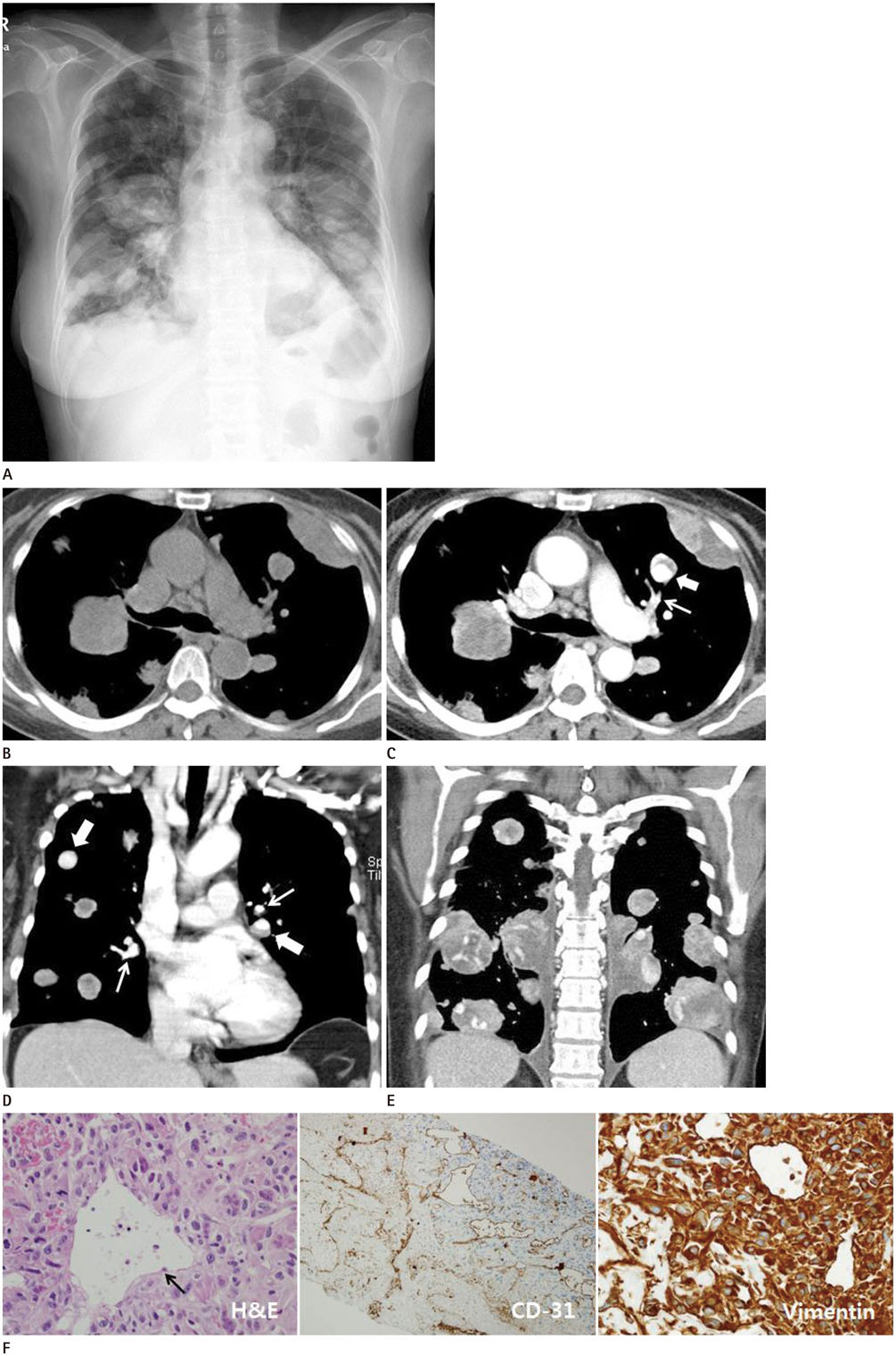
Fig. 1 A 60-year-old woman with pulmonary metastasis from uterine sarcoma, presenting as multiple nodules with tortous, serpentine, aneurysmal dilated intraumor vessels. A. Initial chest radiograph shows multiple, round, variably sized nodules and masses in both lungs, as well as a small amount of pleural effusion. B. Pre-enhanced image shows well-defined homogeneous, variably sized, soft-tissue nodules with a density similar to that of the back muscles and no inner calcific foci. C. Axial contrast-enhanced CT image shows aneurysmal dilatated intratumoral vessel (thick arrow). It has no connection with adjacent branch of the pulmonary artery (thin arrow). D. Coronal contrast-enhanced CT image also show aneurysmal dilatated intratumoral vessels (thick arrows). Branches of the pulmonary artery (thin arrows) pass through the nodules without any connection. E. Coronal contrast-enhanced CT image show multiple variable sized nodues and masses with tortous, dilatated intratumoral or intranodal vessels with an inner cystic or necrotic portion. F. Photomicrographs show tumor cell infiltration with circumscribed cystic spaces lined with flat, endothelial-like cells (arrow), suggesting vascular structures (hematoxylin and eosin, × 100). Cystic spaces show CD-31 positivity (CD-31, × 100) and tumor cells react strongly with mesenchymal antigen (Vimentin, × 400). CD-31 = cluster of differentiation-31, CT = computed tomography
Reference
-
1. Davis SD. CT evaluation for pulmonary metastases in patients with extrathoracic malignancy. Radiology. 1991; 180:1–12.
Article2. Seo JB, Im JG, Goo JM, Chung MJ, Kim MY. Atypical pulmonary metastases: spectrum of radiologic findings. Radiographics. 2001; 21:403–417.
Article3. Daly BD, Cheung H, Gaines PA, Bradley MJ, Metreweli C. Imaging of alveolar soft part sarcoma. Clin Radiol. 1992; 46:253–256.
Article4. Choi JI, Goo JM, Seo JB, Kim HY, Park CK, Im JG. Pulmonary metastases of alveolar soft-part sarcoma: CT findings in three patients. Korean J Radiol. 2000; 1:56–59.
Article5. Shah SH, Jagannathan JP, Krajewski K, O'Regan KN, George S, Ramaiya NH. Uterine sarcomas: then and now. AJR Am J Roentgenol. 2012; 199:213–223.
Article6. Tirumani SH, Deaver P, Shinagare AB, Tirumani H, Hornick JL, George S, et al. Metastatic pattern of uterine leiomyosarcoma: retrospective analysis of the predictors and outcome in 113 patients. J Gynecol Oncol. 2014; 25:306–312.
Article7. Sahdev A, Sohaib SA, Jacobs I, Shepherd JH, Oram DH, Reznek RH. MR imaging of uterine sarcomas. AJR Am J Roentgenol. 2001; 177:1307–1311.
Article8. Ueda M, Otsuka M, Hatakenaka M, Sakai S, Ono M, Yoshimitsu K, et al. MR imaging findings of uterine endometrial stromal sarcoma: differentiation from endometrial carcinoma. Eur Radiol. 2001; 11:28–33.
Article9. Kreibich M, Siepe M, Kroll J, Höhn R, Grohmann J, Beyersdorf F. Aneurysms of the pulmonary artery. Circulation. 2015; 131:310–316.
Article10. Nguyen ET, Silva CI, Seely JM, Chong S, Lee KS, Müller NL. Pulmonary artery aneurysms and pseudoaneurysms in adults: findings at CT and radiography. AJR Am J Roentgenol. 2007; 188:W126–W134.
Article
- Full Text Links
-
- Actions
-
Cited
- CITED
-
- Close
- Share
-
- Similar articles
-
- Pulmonary Metastases of Alveolar Soft-Part Sarcoma: CT Findings in Three Patients
- A Case of Intrapulmonary Lymph Nodes Presenting Multiple Nodules
- A Case of Benign Multiple Pulmonary Nodules in a Patient with Osteosarcoma
- Pulmonary Metastases of Uterine Endometrial Stromal Sarcoma: Diffuse Micronodular and Ground Glass Opacities: A Case Report
- Endometrial Stromal Sarcoma Presented as an Incidental Lung Mass with Multiple Pulmonary Nodules
