Morphometric Study of the Lumbar Posterior Longitudinal Ligament
- Affiliations
-
- 1Department of Neurosurgery, Seoul National University Boramae Medical Center, Seoul, Korea.
- 2Department of Neurosurgery, Soonchunhyang University Hospital Seoul, Seoul, Korea.
- 3Department of Neurosurgery, Gangnam Severance Hospital, Seoul, Korea.
- 4Department of Neurosurgery, Soonchunhyang University Hospital Cheonan, Cheonan, Korea. jwdohns@schca.ac.kr
- KMID: 2403528
- DOI: http://doi.org/10.3340/jkns.2017.0257
Abstract
OBJECTIVE
Morphometric data for the lumbar posterior longitudinal ligament (PLL) was investigated to identify whether there is a difference in the morphometry of the PLL of the lumbar spine at each level with respect to the pattern of intervertebral disc displacement.
METHODS
In 14 formalin-fixed adult cadavers (12 males and 2 females), from L1 to L5, the authors measured the width and height of the PLL and compared them with other landmarks such as the disc and the pedicle.
RESULTS
Horizontally, at the upper margin of the disc, the central portion of the superficial PLL covered 17.8-36.9% of the disc width and the fan-like portion of the PLL covered 63.9-76.7% of the disc width. At the level of the median portion of the disc, the PLL covered 69.1-74.5% of the disc width. Vertically, at the level of the medial margin of the pedicle, the fan-like portion of the PLL covered 23.5-29.9% of the disc height. In general, a significant difference in length was not found in the right-left and male-female comparisons.
CONCLUSION
This study presents the morphometric data on the pattern of intervertebral disc displacement and helps to improve the knowledge of the surgical anatomy of the lumbar PLL.
MeSH Terms
Figure
-
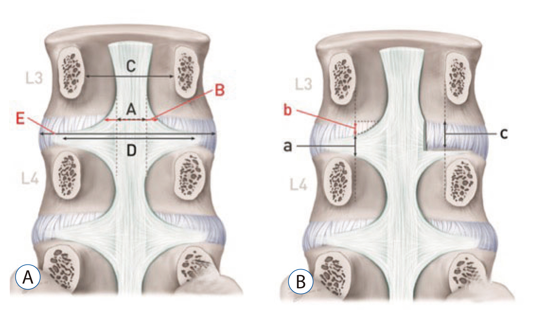
Fig. 1 Parameters. A: Horizontal direction. B: Vertical direction. (A) The width of the central portion of the superficial PLL at the level of the upper margin of the disc. (B) The width of the PLL at the level of the upper margin of the disc. (C) The distance between both pedicles. (D) The width of the PLL at the level of median portion of the disc. (E) The width of the disc at the level of median portion of the disc. (a) The height of the PLL at the level of the medial margin of the pedicle. (b) The height of the disc not covered by the PLL at the level of the medial margin of the pedicle. (c) The disc height at the level of the medial margin of the pedicle. PLL: posterior longitudinal ligament.
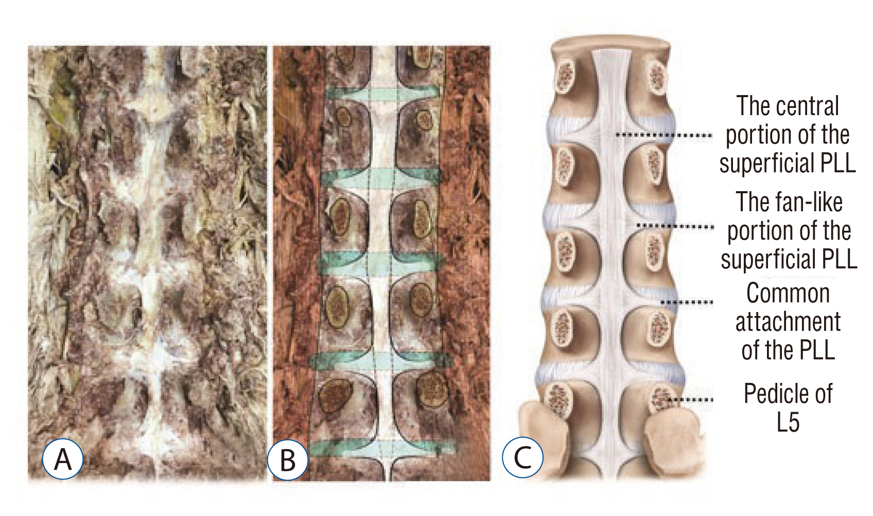
Fig. 2 Photographic (A), drawing (B), and schematic (C) images of the lumbar spine after removing the posterior structure from L1 to L5.
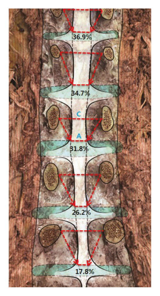
Fig. 3 A/C (%) decreased from L1 to L5. On the contrary, relative horizontal width which is not covered by the central portion of the PLL was increased. Note: most of the herniation of the lumbar intervertebral disc displacement occurs at L4/5, L5/S13,4,5). (A) The width of the central portion of the superficial PLL at the level of the upper margin of the disc. (C) The distance between both pedicles. PLL: posterior longitudinal ligament.

Fig. 4 Schematic (A) and photographic drawing (B) images indicate the anatomical weak point (W) that is not covered by any portion of the PLL. (b) The height of the disc not covered by the PLL at the level of the medial margin of the pedicle. (W) The anatomical weak point regarding herniation of the disc in the spinal canal because it is not covered by any portion of the PLL. PLL: posterior longitudinal ligament.
Reference
-
References
1. Behrsin JF, Briggs CA. Ligaments of the lumbar spine: a review. Surg Radiol Anat. 10:211–219. 1988.
Article2. Bogduk N. Clinical anatomy of the lumbar spine and sacrum. ed 4. Edinburgh: Elsevier Health Sciences;2005. p. 41–42.3. Bosacco SJ, Berman AT. Surgical management of lumbar disc disease. Radiol Clin North Am. 21:377–393. 1983.4. Bosacco SJ, Berman AT, Raisis LW, Zamarin RI. High lumbar disk herniations. Case reports Orthopedics. 12:275–278. 1989.5. Decker HG, Shapiro SW. Herniated lumbar intervertebral disks; results of surgical treatment without the routine use of spinal fusion. AMA Arch Surg. 75:77–84. 1957.6. Ebeling U, Reulen HJ. Are there typical localisations of lumbar disc herniations? A prospective study. Acta Neurochir (Wien). 117:143–148. 1992.
Article7. El-Rakhawy M, El-Shahat AER, Labib I, Abdulaziz E. Lumbar vertebral canal stenosis: concept of morphometric and radiometric study of the human lumbar vertebral canal. Anatomy. 4:51–62. 2010.
Article8. Gray H. Anatomy of the human body. ed 20. Philadelphia: Lea & Febiger;1918.9. Hardy RW, Ball PA. Treatment of disk disease of the lumbar spine. Youmans Neurological Surgery. ed 5. Philadelphia: WB Saunders Company;2004.10. Hsu K, Zucherman J, Shea W, Kaiser J, White A, Schofferman J, et al. High lumbar disc degeneration. Incidence and etiology. Spine (Phila Pa 1976). 15:679–682. 1990.
Article11. Kapoor Y. Morphometry of the lumbar vertebrae and its clinical significance. Sch J App Med Sci. 2:1045–1052. 2014.12. Loughenbury PR, Wadhwani S, Soames RW. The posterior longitudinal ligament and peridural (epidural) membrane. Clin Anat. 19:487–492. 2006.
Article13. Modic MT, Ross JS. Lumbar degenerative disk disease. Radiology. 245:43–61. 2007.
Article14. Ohshima H, Hirano N, Osada R, Matsui H, Tsuji H. Morphologic variation of lumbar posterior longitudinal ligament and the modality of disc herniation. Spine (Phila Pa 1976). 18:2408–2411. 1993.
Article15. Parke WW, Bono CM, Garfin SR. Applied anatomy of the spine. Rothman-Simeone The Spine. ed 6. Philadelphia: Saunders;2011. p. 26–27.16. Prasad R, Hoda MF, Dhakal MM, Singh K, Srivastava A, Sharma V. Epidemiological characteristics of lumbar disc prolapse in atertiary care hospital. Int J Neurosurg. 3:534–536. 2006.17. Prestar FJ. Morphology and function of the interspinal ligaments and the supraspinal ligament of the lumbar portion of the spine. Morphol Med. 2:53–58. 1982.18. Putz R. The detailed functional anatomy of the ligaments of the vertebral column. Ann Anat. 174:40–47. 1992.
Article19. Rosner MK, Campbell VA. Treatment of Disk Disease of the Lumbar Spine Youmans Neurological Surgery. ed 6. Philadelphia: WB Saunders Company;2011. p. 2919.20. Santiago FR, Milena GL, Herrera RO, Romero PA, Plazas PG. Morphometry of the lower lumbar vertebrae in patients with and without low back pain. Eur Spine J. 10:228–233. 2001.
Article21. Wiltse LL. Anatomy of the extradural compartments of the lumbar spinal canal. Peridural membrane and circumneural sheath. Radiol Clin North Am. 38:1177–1206. 2000.
- Full Text Links
-
- Actions
-
Cited
- CITED
-
- Close
- Share
-
- Similar articles
-
- Ossification of the Posterior Longitudinal Ligament: 2 cases report
- Intradural Lumbar Disc Herniations Associated with Epidural Adhesion : Report of Two Cases
- Cauda Equina Syndrome due to Lumbar Ossification of the Posterior Longitudinal Ligament: A Case Report
- Ganglion and Synovial Cyst on the Posterior Longitudinal Ligament: Case Report
- Anterior Decompression and Interbody Fusion for Ossification of the Posterior Longitudinal Ligament of the Cervical Spine: Case Report
