Periductal Stromal Sarcoma of the Breast: a Case Report
- Affiliations
-
- 1Department of Radiology, Dankook University Hospital, Cheonan, Korea. sirenos@hanmail.net
- KMID: 2400382
- DOI: http://doi.org/10.13104/imri.2017.21.4.269
Abstract
- Periductal stromal sarcoma (PSS) is a type of rare malignant fibroepithelial tumor. PSS is a recently introduced diagnostic entity and there are few reports about radiological features of this tumor. Pre-operative diagnosis is difficult because it reveals similar symptoms with other benign and malignant tumors with absence of specific radiologic findings. We present a woman age 30 that underwent mammotome biopsy for a BI-RADS 4 lesion on her left breast and received histopathology diagnosis of a phyllodes tumor. Additionally, she underwent a wide excision depending on her histopathology diagnosis. Her final diagnosis was PSS. Six months later, no recurrence was detected. However, frequent follow-up is needed because PSS can develop into phyllodes tumor or entity of breast cancer.
MeSH Terms
Figure
-
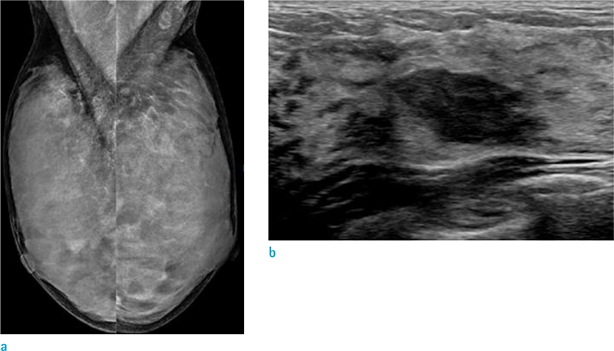
Fig. 1 (a) Mammogram mediolateral oblique (MLO) view reveals extremely dense breasts that make it difficult to evaluate the mass lesion. (b) Ultrasonographic image reveals oval mass in parallel orientation with angular margins in upper outer quadrant of the left breast.
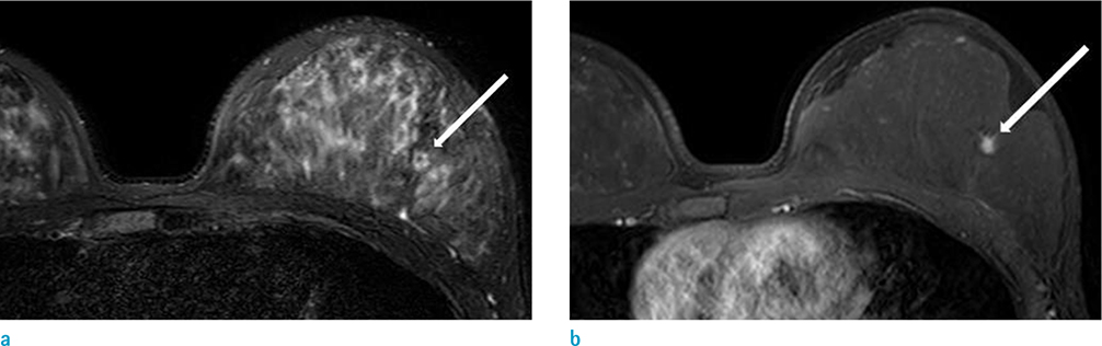
Fig. 2 On MRI of breasts after mammotome excision, a 6 mm-sized irrgular shaped mass (arrow), revealing high signal intensity on fat-saturated T2-weighted image (a) with homogeneous enhancement (arrow) on first dynamic enhancement study (90 seconds) (b) was found in upper outer quadrant of the left breast.
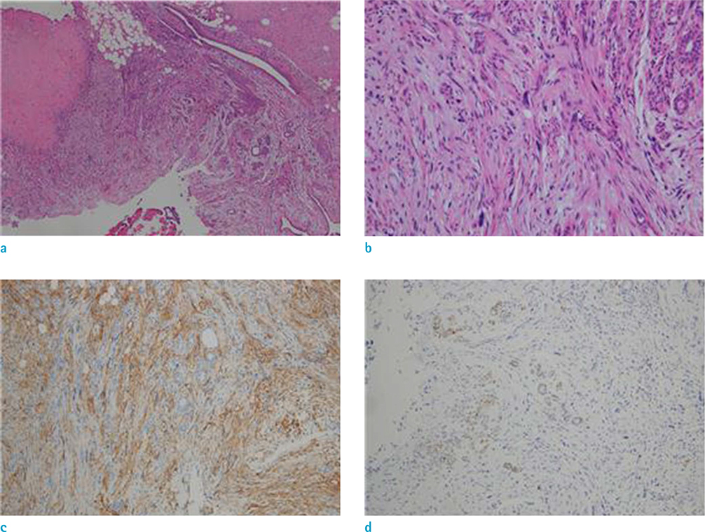
Fig. 3 On pathology examination, (a) low-power field (× 40) and (b) high-power field (× 200) images of the tumor reveal biphasic proliferation and a multinodular growth pattern and was composed of spindle cells without significant atypia, cuff formations around open tubules without leaf-like pattern. (c) On immunohistochemistry staining, stromal cells were positive with CD34 and (d) negative with S100.
Reference
-
1. Tavassoli FA, Devilee P. Pathology and genetics tumours of the breast and female genital organs. In : Tavassoli FA, Devilee P, editors. World Health Organization classification of tumours. Lyon: IARC Press;2003. p. 101–102.2. Burga AM, Tavassoli FA. Periductal stromal tumor: a rare lesion with low-grade sarcomatous behavior. Am J Surg Pathol. 2003; 27:343–348.3. Rao AC, Geetha V, Khurana A. Periductal stromal sarcoma of breast with lipoblast-like cells: a case report with review of literature. Indian J Pathol Microbiol. 2008; 51:252–254.
Article4. Pandey M, Mathew A, Abraham EK, Rajan B. Primary sarcoma of the breast. J Surg Oncol. 2004; 87:121–125.
Article5. Callery CD, Rosen PP, Kinne DW. Sarcoma of the breast. A study of 32 patients with reappraisal of classification and therapy. Ann Surg. 1985; 201:527–553.6. Oberman HA, Nosanchuk JS, Finger JE. Periductal stromal tumors of breast with adipose metaplasia. Arch Surg. 1969; 98:384–387.
Article7. Tomas D, Jankovic D, Marusic Z, Franceschi A, Mijic A, Kruslin B. Low-grade periductal stromal sarcoma of the breast with myxoid features: Immunohistochemistry. Pathol Int. 2009; 59:588–591.
Article8. Masbah O, Lalya I, Mellas N, et al. Periductal stromal sarcoma in a child: a case report. J Med Case Rep. 2011; 5:249.
Article9. Tse GM, Tan PH, Lui PC, Putti TC. Spindle cell lesions of the breast--the pathologic differential diagnosis. Breast Cancer Res Treat. 2008; 109:199–207.
Article10. Hungermann D, Buerger H, Oehlschlegel C, Herbst H, Boecker W. Adenomyoepithelial tumours and myoepithelial carcinomas of the breast--a spectrum of monophasic and biphasic tumours dominated by immature myoepithelial cells. BMC Cancer. 2005; 5:92.
Article
- Full Text Links
-
- Actions
-
Cited
- CITED
-
- Close
- Share
-
- Similar articles
-
- Mammographic and Sonographic Findings of Periductal Mastitis: A Case Report
- Endometrial Stromal Sarcoma Presented as a Multilocular Cystic Mass without a Solid Component: A Case Report
- Two Cases of Low Grade Endometrial Stromal Sarcoma
- A Case of High-grade Endometrial Stromal Sarcoma with Metastasis to the Stomach
- A Case of Low-Grade Endometrial Stromal Sarcoma of the Uterus (So-Called ""Endolymphatic Stromal Myosis"")
