Progressive Transformation of Germinal Centers in Presacral Space: MRI Findings and Literature Review
- Affiliations
-
- 1Department of Radiology, Anam Hospital, Korea University College of Medicine, Seoul, Korea. urorad@korea.ac.kr
- 2Department of Pathology, Anam Hospital, Korea University College of Medicine, Seoul, Korea.
- KMID: 2376271
- DOI: http://doi.org/10.13104/imri.2017.21.1.56
Abstract
- Progressive transformation of germinal centers (PTGC) is an atypical feature seen in lymph nodes with unknown pathogenesis. PTGC most commonly presents in adolescent and young adult males as solitary painless lymphadenopathy with various durations. Cervical nodes are the most commonly involved ones while involvements of axillary and inguinal nodes are less frequent. PTGC develops extremely rarely in other locations. We report a rare case of solitary mass present in the presacral space. The mass as subsequently proven to be PTGC. To the best of our knowledge, PTGC in the presacral space has not been previously reported in the literature.
MeSH Terms
Figure
-
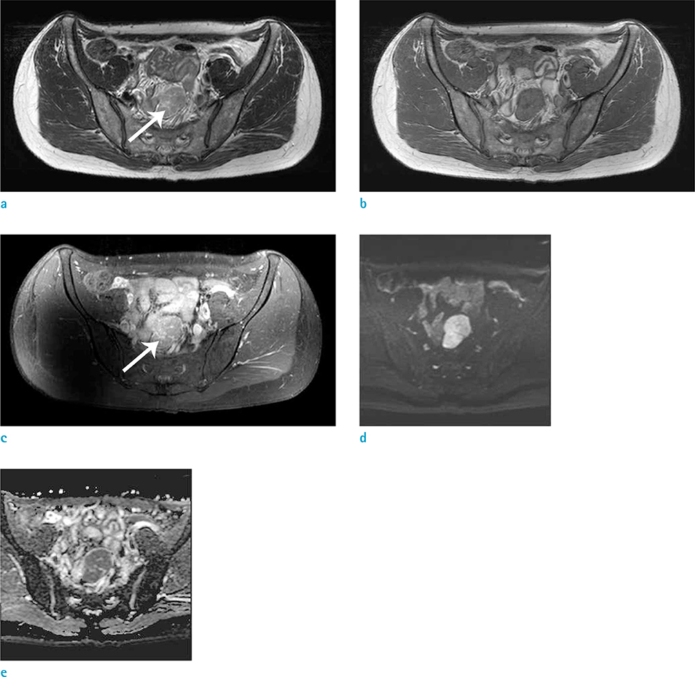
Fig. 1 Axial T2-weighted turbo spin-echo (a) and axial T1-weighted turbo spin-echo (b) MR images showing a well-circumscribed presacral mass with slightly high signal intensity on T2-weighted imaging and iso-signal intensity on T1-weighted imaging. Axial contrast-enhanced T1-weighted subtraction MRI (c) showing homogeneous enhancement on 1-min delayed phase. DWI (b-value = 800 s/mm2) (d) showing high signal intensity in the mass. (e) The mass demonstrates low signal intensity on ADC map. A few small high T2 signal intensity foci (a, arrow) showing strong contrast enhancement (c, arrow) were scattered in the center of the mass. These foci were not shown on DWI or ADC map due to their small sizes.
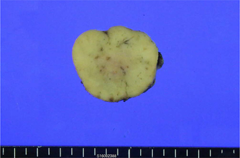
Fig. 2 The specimen with a round shaped soft tissue measured at 3.6 × 3.3 × 1.8 cm. The external surface is smooth. On section, the cut surface shows a diffusely yellowish-gray homogeneous appearance.
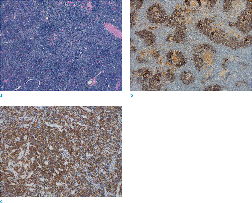
Fig. 3 The section from lymph nodes with variable sizes and enlarged follicles with indistinct margins. It is composed of follicular mantle lymphocytes and extensive follicular dendritic cells. H&E stain, × 40 (a). Positive CD21 immunostaining of follicular dendritic cells, × 40 (b). Bcl-2-positive mantle zone B cells infiltrate and disrupt germinal centers, × 200 (c).
Reference
-
1. Lennert K, Muller-Hermelink HK. Lymphocytes and their functional forms - morphology, organization and immunologic significance. Verh Anat Ges. 1975; 69:19–62.2. Leong A. A pattern approach to lymph node diagnosis. New York: Springer;2011.3. Hicks J, Flaitz C. Progressive transformation of germinal centers: review of histopathologic and clinical features. Int J Pediatr Otorhinolaryngol. 2002; 65:195–202.4. Park SW, Jang SM, Kim DY, Son JH, Cho YC, Sung IY. Progressive transformation of germinal centers in submandibular area. J Korean Assoc Maxillofac Plast Reconstr Surg. 2011; 33:368–372.5. Miller MW, Gatter KM, Cannady SB, Wax MK. Progressive transformation of germinal centers (PTGC) in the head and neck. Laryngoscope. 2010; 120:Suppl 4. S168.6. Chang CA, Kumar B, Nandurkar D. A case report of high 18F-FDG PET/CT uptake in progressive transformation of the germinal centers. Medicine (Baltimore). 2015; 94:e412.7. Hain KS, Pickhardt PJ, Lubner MG, Menias CO, Bhalla S. Presacral masses: multimodality imaging of a multidisciplinary space. Radiographics. 2013; 33:1145–1167.8. Bonekamp D, Horton KM, Hruban RH, Fishman EK. Castleman disease: the great mimic. Radiographics. 2011; 31:1793–1807.9. Pickhardt PJ, Bhalla S. Unusual nonneoplastic peritoneal and subperitoneal conditions: CT findings. Radiographics. 2005; 25:719–730.
- Full Text Links
-
- Actions
-
Cited
- CITED
-
- Close
- Share
-
- Similar articles
-
- Progressive Transformation of Germinal Centers in Axillary Lymph Nodes Mimicking Metastatic Lymphadenopathy after Breast Cancer Surgery: A Case Report
- Progressive Transformation of Germinal Centers in Submandibular Area: Case Report
- A Case of Presacral Cystic Teratoma
- Laparoscopic Resection of Presacral Tumor: A New Approach in the Era of the Minimally Invasive Surgery
- Clinical Course of Myasthenic Patients after Thymectomy and Relationship between Germinal Center and Postoperative Course
